Comparative Guts

Search
Close this search box.

1600 CE

An example of 17th-century Sino-European Cross-cultural medical history

Marta Hanson

Independent scholar, MPIWG Affiliate
Introduction
These five images are all concerned with the work of specific Jesuits to introduce Chinese medicine to Europeans in the second half of the seventeenth century, in terms of bringing to Europe Chinese medical texts [Images 1, 2] and reproducing body images in them [Images, 3, 4, 5]. The earliest record of Chinese images of the human body moving west started over 300 years earlier with the Persian polymath, physician, and historian, Rashīd al-Dīn (1247–1318), who commissioned a Persian translation of Chinese medical texts. The resulting Tānksūqnāma-yi Īl-khān dar funūn-i ulūm-i khatāyī  (The Treasure Book of the Ilkhān  on Chinese Science and Techniques, 1313: Tansūqnāma  for short) is partly extant today in one manuscript at the Aya Sofya Library in Istanbul. There is no extant evidence, however, of continued engagement with anatomical images in either direction, or further West, until the seventeenth century. Jesuits were involved in translating European anatomy into Chinese, starting with Johann Schreck’s (1576-1630) Abstract of the Western Theory of the Human Body  (Taixi renshen shuogai, 1625), and even into Manchu by the 1720s. Jesuits also translated Chinese body images into Latin in the Specimen medicinæ sinicæ (A Sample of Chinese Medicine, 1682). Zhang Jiebin’s Classified Canon, Illustrated Wing  (1624) was the main Chinese source text for the Specimen’s 
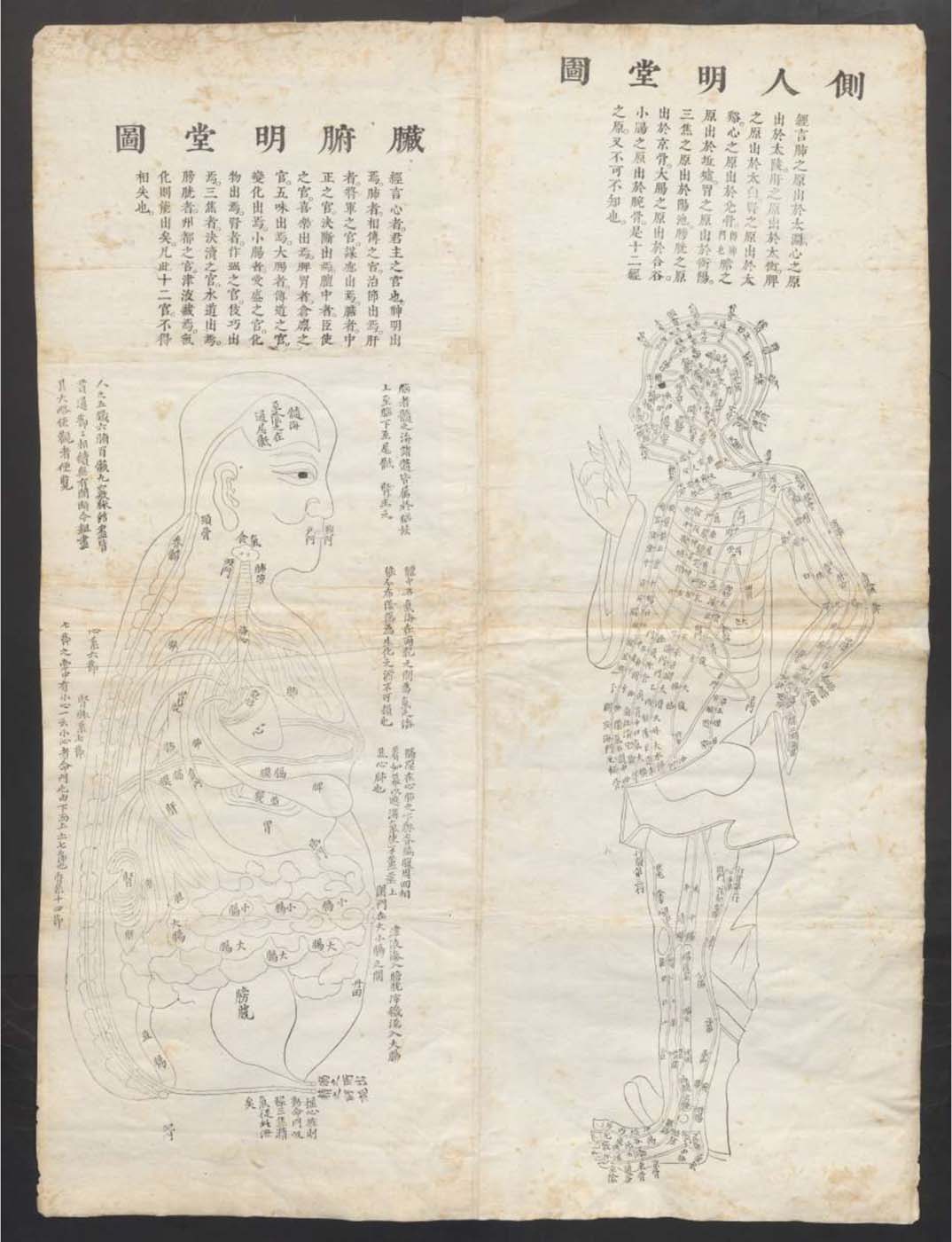
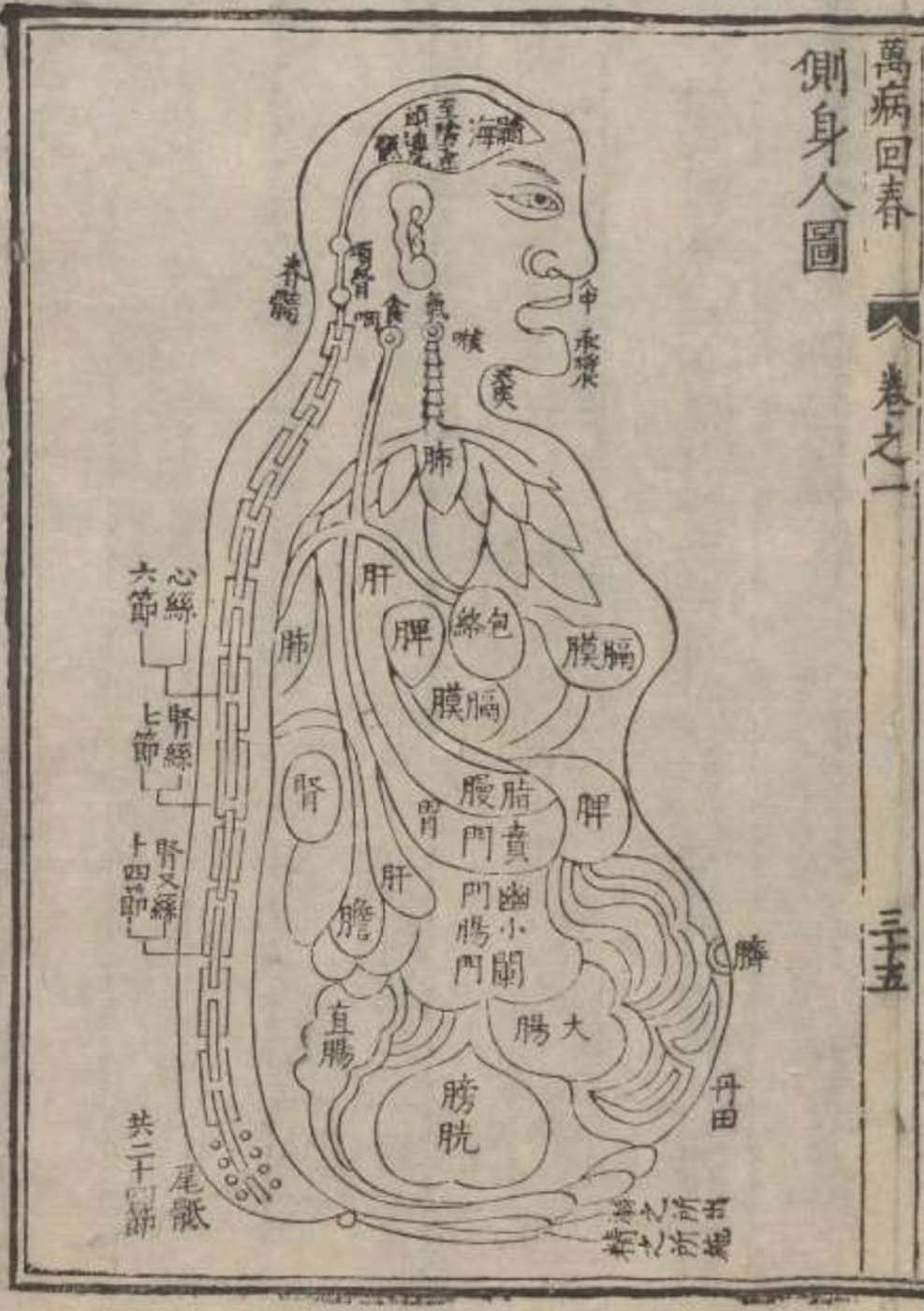
images of Chinese medical concepts of twelve viscera-channels.  The two images of the Chinese “viscera man” (images 1 & 
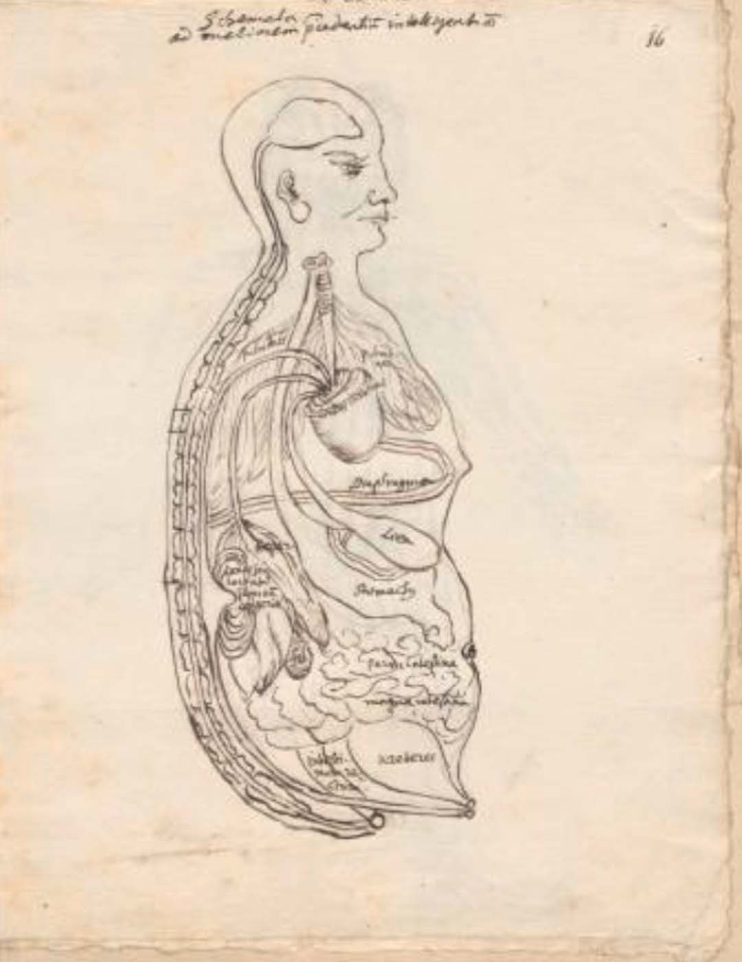
2), however, come from two other medical texts that the Flemish Jesuit Philippe Couplet (1623-1693) may have given to the German Christian Mentzel (1622-1701), personal physician to the Great Elector Friedrich Wilhelm of Brandenberg (1620-1688) and curator of his Chinese collection, who was interested in Chinese medicine. Mentzel , in fact, appears to have also drawn the Chinese “viscera man” 
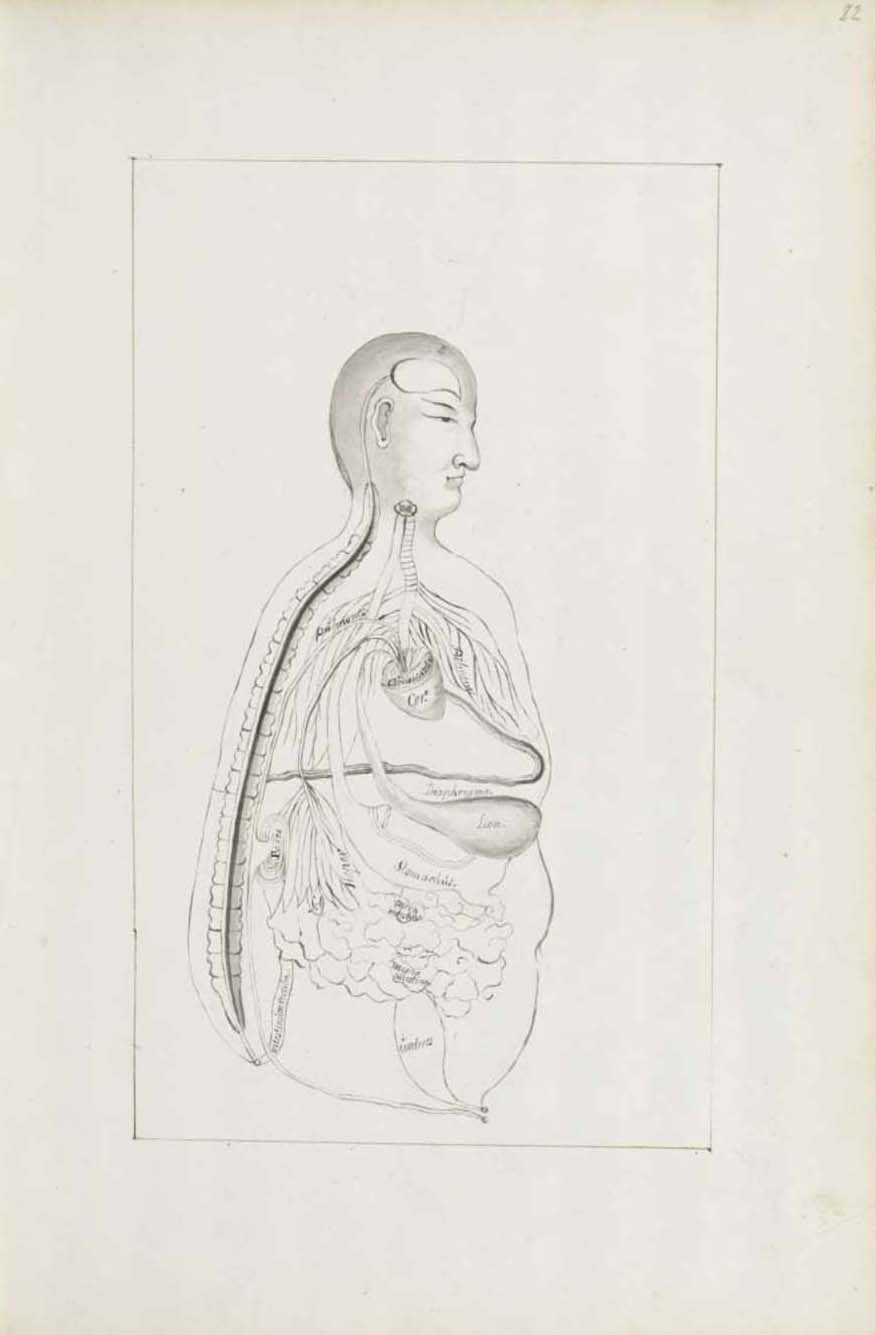
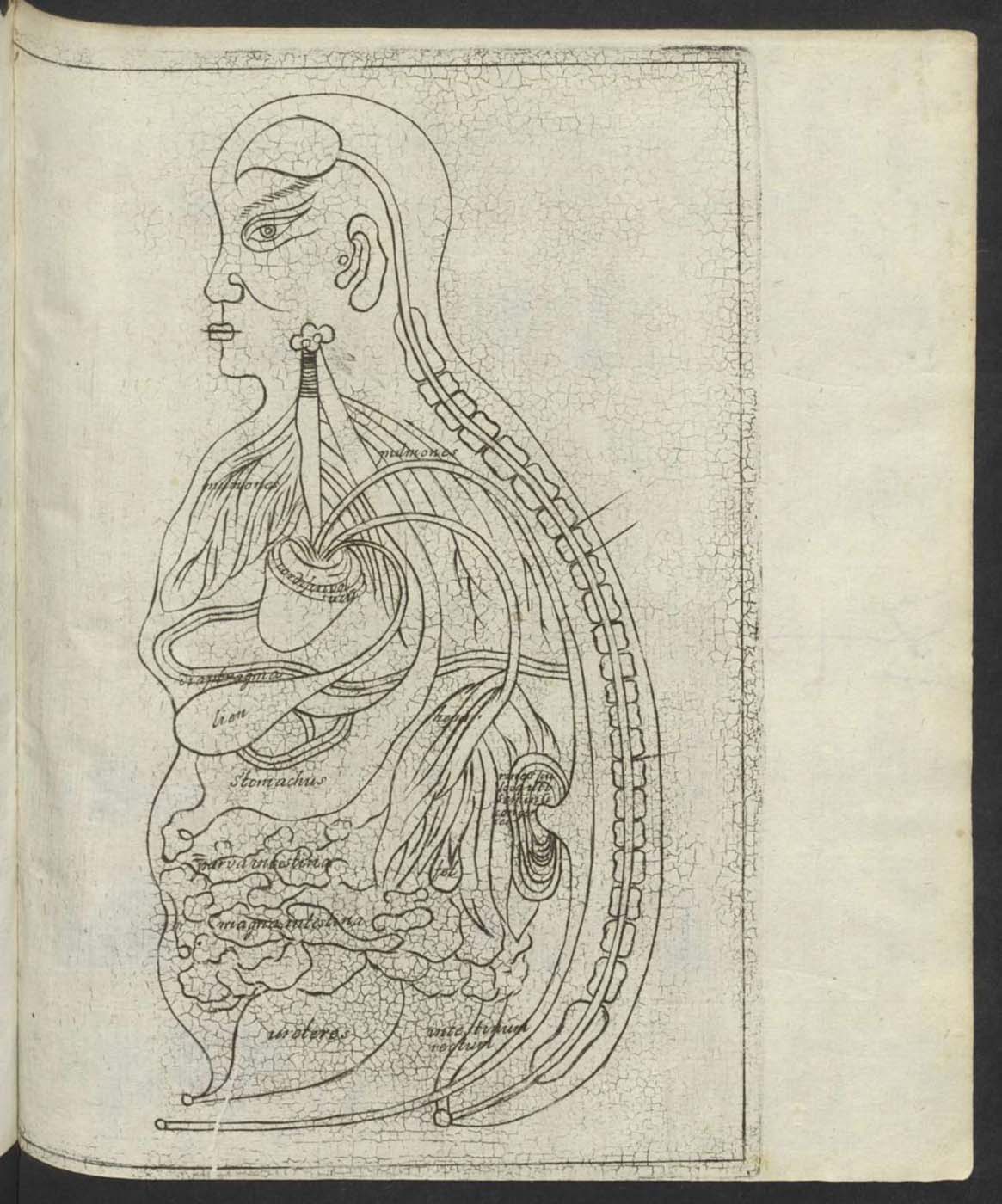
(image 3) as means to learn Chinese concepts of human viscera depicted as well in the manuscript (image 4) and printed (image 5) versions of Specimen medicinæ sinicæ. Chinese conceptions of a four-valve heart and the reproductive role of kidneys were successfully translated (fig. 3, 4, 5), despite differing from contemporary European understandings of the heart and kidneys. The Chinese guts, by contrast, are visually and textually commensurable with European understandings of guts being located in the lower abdomen and having a transporting role.
Guts in 17th-century Sino-European medical history

The Chinese debated what viscera were contained with the human body, where they were located, and what functions they carried out. Various depictions of the “viscera man,” a conventional side-body view of a “see-through” human body with head and viscera but no limbs, provide evidence of these debates. In one version (fig. 1), a three-valve heart dominates the upper section with a valve-tube going (from top down) to the kidneys, liver, and spleen. Another viscera-man from the same period (fig. 2), by contrast, depicts a one-valve heart. Furthermore, the guts dominate the lower part of all “viscera man” images, but they are depicted and discussed differently. The guts could be drawn like floating clouds across the entire abdomen (fig. 1) or resemble a large sac between the small intestine above and the bladder below (fig. 2).

Different types of metaphors were also deployed to explain visceral functions. Whereas the bureaucratic metaphor dominated in some descriptions of the “viscera man” (fig. 1), the hydraulic metaphor dominated in other ones (fig. 2). Although different metaphors are not obvious in different “viscera man” images, they nonetheless informed different emphasizes in textual descriptions. The bureaucratic metaphor portrayed the guts as the ministry of transportation through which transformed substances moved (fig. 1). The hydraulic metaphor, however, emphasized the course of the internal tract that links the large intestine with the lungs in a yin-yang pair and the guts’ materiality (fig. 2). In the average human, the guts weigh two jin  (1 jin=0.5 kilo) and 12 liang  (1 liang = 50 g) and are two zhuang and one chi long (1 zhuang=3.3 meters, 1 chi=0.33 meters), two and a half cun wide (1 cun=3.33 centimetres) wide, and eight fen in diameter (1 fen=0.33 centimetres).

Remarkably, the three earliest known European interpretations of the Chinese “viscera man” are all extant today and are related to Specimen medicinæ sinicæ (1682), the first printed Latin translation of Chinese medicine exemplifying Sino-European cross-cultural exchange. Whereas Christian Mentzel (1622-1701), personal physician to the Great Elector Friedrich Wilhelm of Brandenberg (1620-1688) and curator of his Chinese collection, drew one sketch (fig. 3), the artists of the other two drawings, one (fig. 4), in the only known manuscript of Specimen, and one (fig. 5), in the printed Specimen, remain unknown. All three sketches preserve Chinese medical conceptions depicted in the Chinese source (fig. 1). The three-valve heart did not align, for example, with the four-valve heart understood in Europe since Harvey’s 1628 publication on the heart’s role in the circulation of the blood (fig. 3, 4, 5). Mentzel also recorded the Chinese conception of the kidney’s role in reproduction – “Kidneys, or the sites where the mass of the seeds (or semen) are” – in Latin “Renes seu loca ubi seminum congeries” (fig. 3). He took the Latin from the printed Specimen’s “viscera man” (fig. 5). By contrast, the Latin phrases – parva intestina and magna intestina (fig. 3, 4, 5) – clearly designate the cloud-like formations across the abdomen as the small and large intestines situated between the stomach (Stomachus) above and bladder (ureteres) below. At this level of abstraction, the guts appear to have not needed any further translation.

BIBLIOGRAPHY
Hanson, M. and Pomata, G. “A Sample of Chinese Medicine for 17th-Century Europe: SBB-Berlin Ms. lat. fol. 95.” In “Sammellust und Wissensdrang – Vier Jahrhunderte Asiatica in Berlin” (“Connoisseurial Passion and Thirst for Knowledge – Four centuries of Asiatica in Berlin”). Publication by the East Asia Department, Staatsbibliothek zu Berlin, dedicated to its Triple Jubilee 400 years Christian Mentzel – 100 years East Asia Department – 70 years German Science Foundation funding. Berlin: forthcoming 2023.
Hanson, M. and Pomata, G. “A Glimpse into the Making of Specimen Medicinae Sinicae: SBB-Berlin Ms. sin. 11.” In “Sammellust und Wissensdrang – Vier Jahrhunderte Asiatica in Berlin” (“Connoisseurial Passion and Thirst for Knowledge – Four Centuries of Asiatica in Berlin”). Publication by the East Asia Department, Staatsbibliothek zu Berlin, dedicated to its Triple Jubilee 400 years Christian Mentzel – 100 years East Asia Department – 70 years German Science Foundation funding. Berlin: forthcoming 2023.