Ca. 1660s-1681c. Lat. Fol. 95, unknown artist
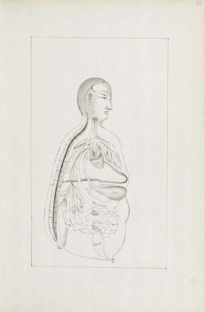
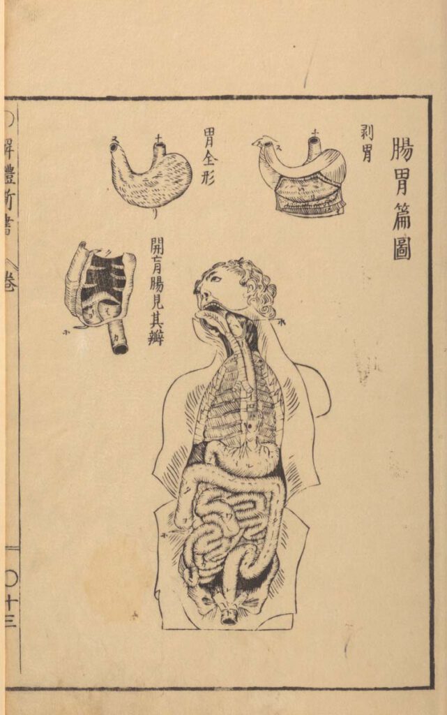
Ca. 1660s-1681c. Lat. Fol. 95, unknown artist This sketch of the “Viscera Man” was also based on a Chinese source (image 1), however, the artist remains anonymous. It was included in the only known extant manuscript (Ms. Lat. Fol. 95 preserved in the Staatsbibliothek in Berlin) of the 1682 printed Specimen Medicinae Sinicae. It illustrates […]
Ca. 1681 Ms sin. 11, part 3 by Christian Mentzel
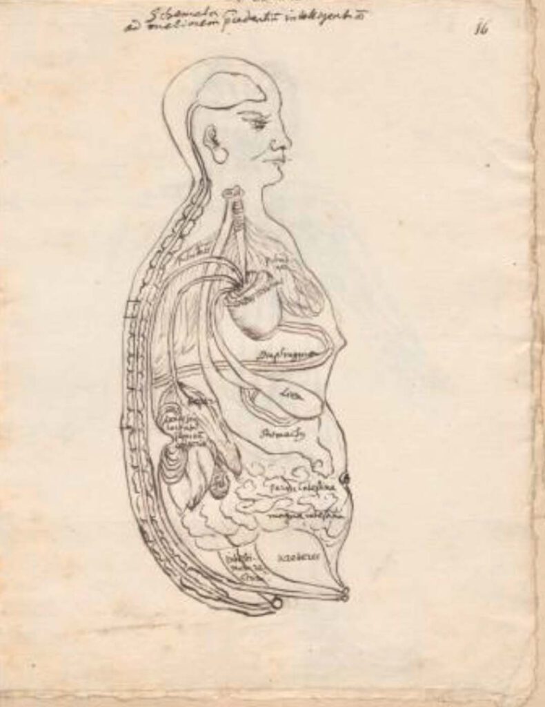
Ca. 1681 Ms sin. 11, part 3 by Christian Mentzel The personal physician to the Great Elector Friedrich Wilhelm of Brandenberg and curator of his library, Christian Mentzel (1622-1701), sketched this “Viscera Man” based on a Chinese source (Image 1). Both Mentzel’s sketch (Image 3) and the printed 1597 original (Image 1) are included in […]
1597 reprint of Wanbing huichun “Diagram of Man’s Side-Body”
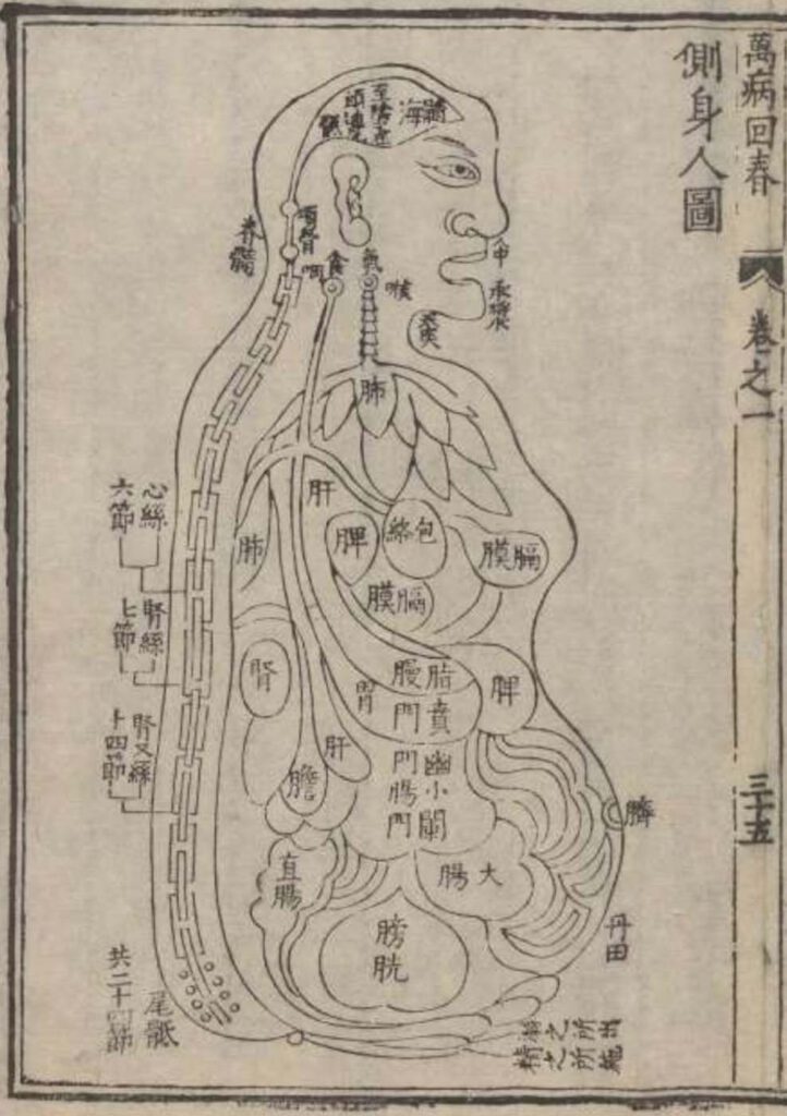
‘Diagram of Man’s Side-Body’ in 1597 reprint of Wanbing huichun (Ceshen ren tu 側身人圖). This version of the “Viscera Man” was printed in a 1597 reprint of the Chinese medical text Myriad diseases ‘Spring Returned’ (i.e., ‘cured’) (Wanbing huichun 萬病回春) that is preserved in the Staatsbibliothek in Berlin. – This 1597 reprint of a “viscera […]
Published in 1597 reprint of a 1565 Yixue gangmu, preserved in Ms sin. 11, part 1, “Enlightened-Hall Diagram of the Viscera”
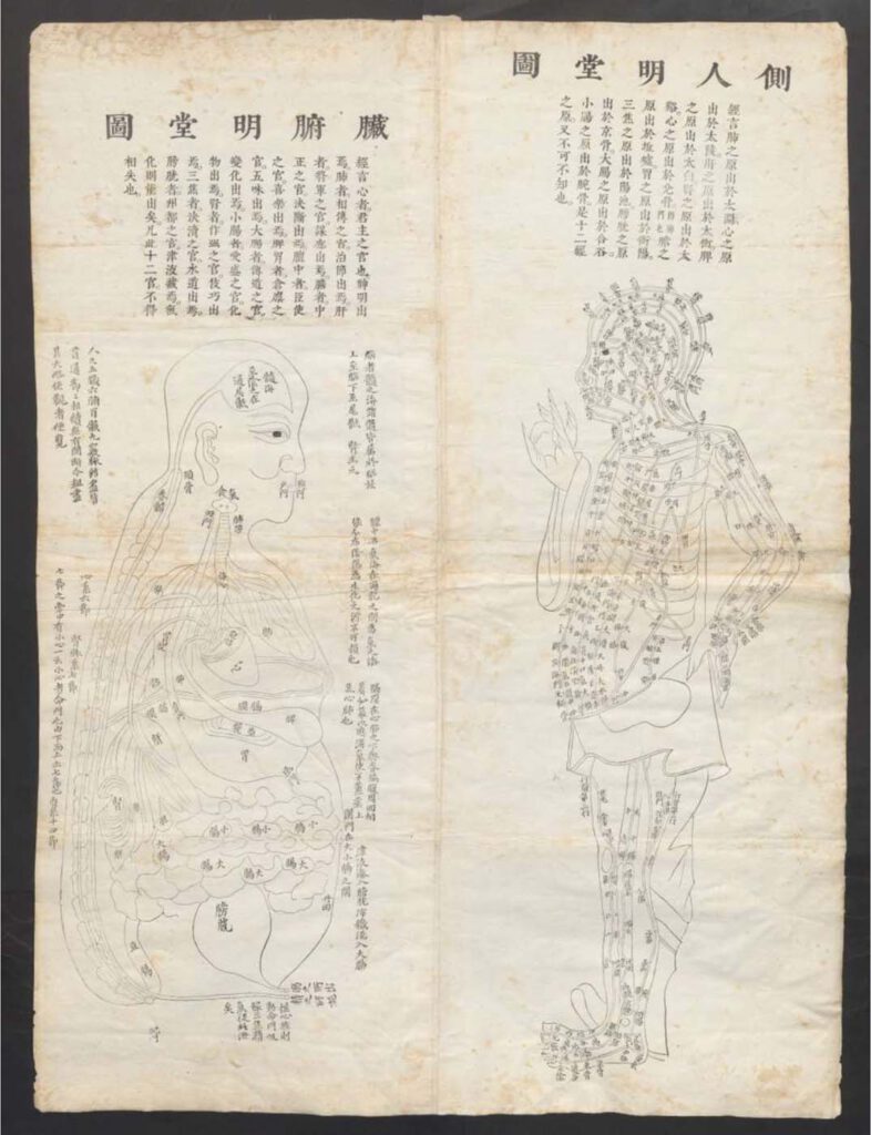
‘Enlightened-Hall Diagram of the Viscera’, in 1597 reprint of a 1565 Yixue gangmu, preserved in Ms sin. 11, part 1 (Zangfu Mingtang tu 臟腑明堂圖). This fold-out plate (ca. 79cm x 58cm) of a “Viscera Man” has been inserted into a manuscript (ms. sin. 11) preserved in Biblioteka Jagiellońska Kraków, Poland, digitalized by the Staatsbibliothek in […]
Sugita Gempaku’s Kaitai shinsho
Sugita Gempaku’s Kaitai shinsho (解体新書, “New Book of Anatomy”; 1774) – Sugita Gempaku undertook his epoch-making translation of a Dutch anatomical manual before he could read Dutch. This point is key: What persuaded him of the manual’s truth was not its text, which he initially couldn’t comprehend, but rather, its images, and more specifically their […]
Reproduced artwork detailing small and large intestines with Tibetan and Latin labels
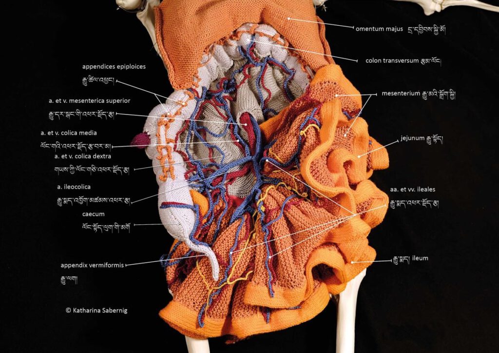
Reproduced artwork detailing small and large intestines with Tibetan and Latin labels Reproduced artwork detailing small and large intestines with Tibetan and Latin labels. Contemporary knitwork. © Katharina Sabernig – Tibetan classical medical vocabulary is already rich in anatomical terms. When encountering biomedical concepts, new terms were coined in the Tibetan language. Nevertheless, many historical […]
Knitted artwork of guts based on Tibetan medical painting at Atsagat Monastery, Republic of Buryatia, Russia.
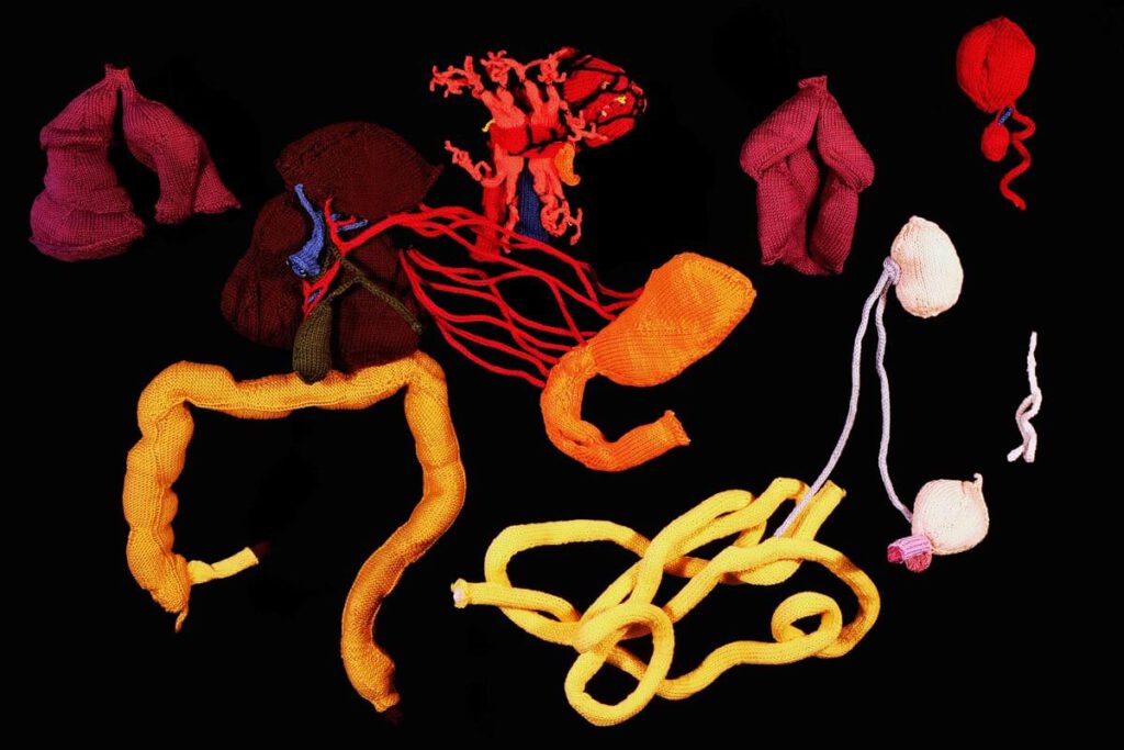
Knitted artwork of guts based on Tibetan medical painting at Atsagat Monastery, Republic of Buryatia, Russia Knitted artwork of guts based on Tibetan medical painting at Atsagat Monastery, Republic of Buryatia, Russia. © Katharina Sabernig – In Buryatia, an ethnically Mongolian region belonging to Russia, Tibetan medical knowledge encountered biomedical knowledge in the early twentieth […]
Mun (Mursi) belly painting

Mun (Mursi) belly painting Mun (Mursi) belly painting Photographer: Kate Fayers-Kerr – Mun Mursi belly painting: the Mun (Mursi) use mud and clay as a technique for grounding self and society in social, political, and cosmological space and order. Fayers-Kerr has persuasively shown how Mun painting and ideas about eating earth are closely connected to […]
Food’s Course of Movement in the Intestines (engraving)
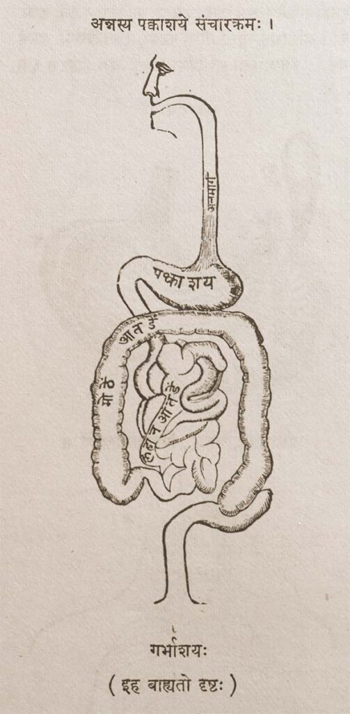
Food’s Course of Movement in the Intestines (engraving) Provenance: Printed Book, page 44. Anglicized Bibliographic data The Śārṅgadhara-saṃhitā by Paṇḍit Śārṅgadharāchārya, son of Paṇḍit Dāmodara with the commentary Aḍhamalla’s Dīpikā and Kāśīrāma’s Gūḍhārtha-Dīpikā. Ed. Paṇḍitaparaśurāmaśāstri. Bombay: Pāndurang Jāwajī, Nirṇaya-Sāgar Press, 1931. Sanskrit Title and Author in Roman font śrīmatpaṇḍitadāmodarasūnu-śārṅgadharācāryaviracitā . śārṅgadharasaṃhitā . bhiṣagvarāḍhamallaviracitadīpikā-paṇḍitakāśirāmavaidyaviracita-gūḍhārthadīpikābhyāṃ ṭīkābhyāṃ saṃvalitā […]
Indian anatomical painting
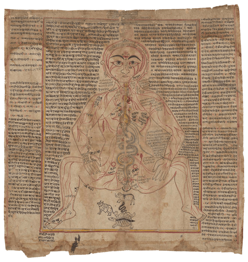
Indian anatomical painting Circa eighteenth century, western India. An old-Gujarati manuscript (circa 1900?). (Wellcome MS Indic d 74. Photo Wellcome Library, London.) Source: Wellcome Collection CC BY-NC-SA 4.0 – Premodern anatomical images in India were influenced by Persian drawings on anatomy, particularly during the Mughal dynasty’s reign in north India from the sixteenth to eighteenth […]