1682 Specimen Medicinae Sinicae, unknown artist
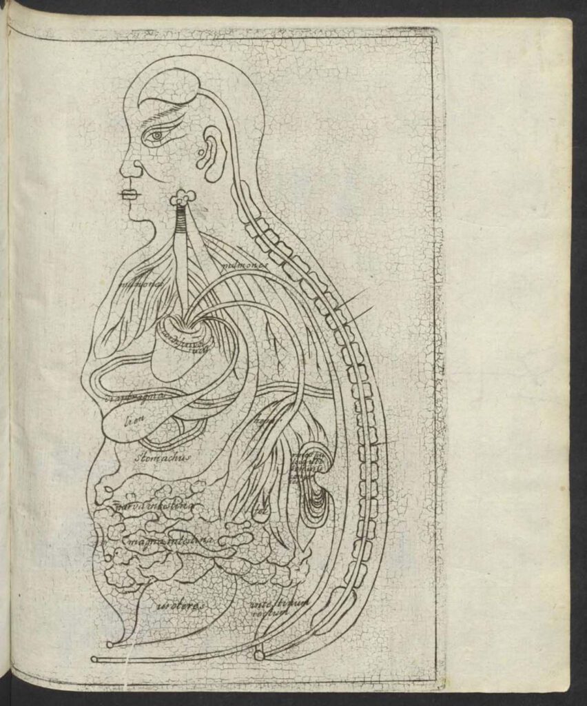
1682 Specimen Medicinae Sinicae, unknown artist This “Viscera Man” was also based on a Chinese source (Image 1) but was printed in the 1682 Specimen Medicinae Sinicae. The anonymous artist retained the Chinese original’s flat rendering (Image 1) rather than the three-dimensional rendering of Christian Mentzel’s sketch (Image 3) and the sketch (Image 4) in […]
Ca. 1660s-1681c. Lat. Fol. 95, unknown artist
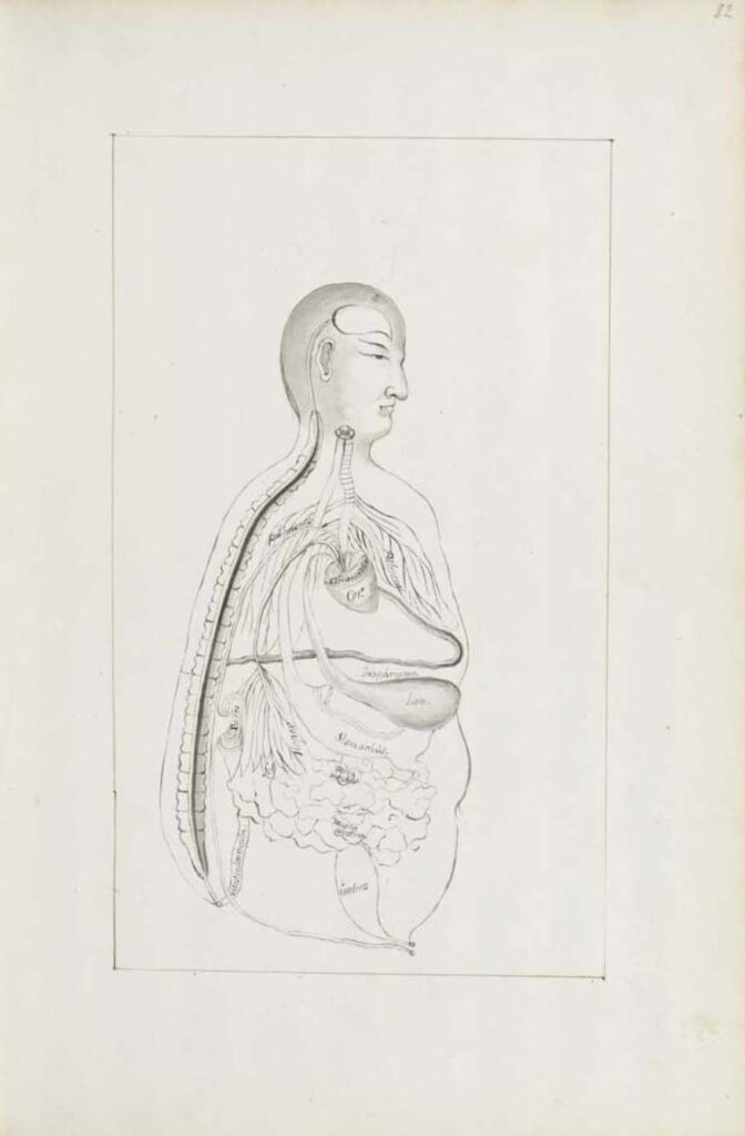
Ca. 1660s-1681c. Lat. Fol. 95, unknown artist This sketch of the “Viscera Man” was also based on a Chinese source (image 1), however, the artist remains anonymous. It was included in the only known extant manuscript (Ms. Lat. Fol. 95 preserved in the Staatsbibliothek in Berlin) of the 1682 printed Specimen Medicinae Sinicae. It illustrates […]
Ca. 1681 Ms sin. 11, part 3 by Christian Mentzel
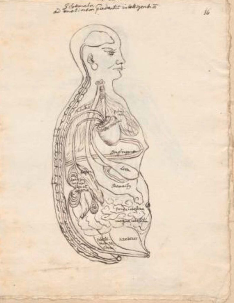
Ca. 1681 Ms sin. 11, part 3 by Christian Mentzel The personal physician to the Great Elector Friedrich Wilhelm of Brandenberg and curator of his library, Christian Mentzel (1622-1701), sketched this “Viscera Man” based on a Chinese source (Image 1). Both Mentzel’s sketch (Image 3) and the printed 1597 original (Image 1) are included in […]
1597 reprint of Wanbing huichun “Diagram of Man’s Side-Body”
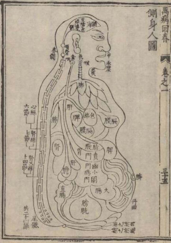
‘Diagram of Man’s Side-Body’ in 1597 reprint of Wanbing huichun (Ceshen ren tu 側身人圖). This version of the “Viscera Man” was printed in a 1597 reprint of the Chinese medical text Myriad diseases ‘Spring Returned’ (i.e., ‘cured’) (Wanbing huichun 萬病回春) that is preserved in the Staatsbibliothek in Berlin. – This 1597 reprint of a “viscera […]
Published in 1597 reprint of a 1565 Yixue gangmu, preserved in Ms sin. 11, part 1, “Enlightened-Hall Diagram of the Viscera”
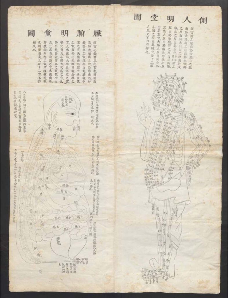
‘Enlightened-Hall Diagram of the Viscera’, in 1597 reprint of a 1565 Yixue gangmu, preserved in Ms sin. 11, part 1 (Zangfu Mingtang tu 臟腑明堂圖). This fold-out plate (ca. 79cm x 58cm) of a “Viscera Man” has been inserted into a manuscript (ms. sin. 11) preserved in Biblioteka Jagiellońska Kraków, Poland, digitalized by the Staatsbibliothek in […]