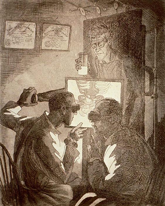
John Sloan, A self-portrait of the artist holding a cup of barium and undergoing an upper gastrointestinal fluoroscopic study under the care of two radiologists, 1926.
Medium: etching and aquatint. Collection: National Library of Medicine.
–
Anatomy is a science predominantly based on visual observation. Therefore, the anatomical body’s existence is seemingly reliant on modes of visual communication—which are constantly, and rapidly, evolving. However, with each new technology used in our seemingly relentless visual interrogation of the body we are not only seeing more of the body, but we are also seeing it differently. Indeed, the anatomical body is as much defined by the methods used to behold it as it is defined by the structures that it claims to reveal.
In the radiography room, which is an anatomy theatre of sorts, the relations and positions of the traditional protagonists are altered. The observed body is no longer necessarily a dead body, but is alive and conscious.
A comparison can be made here with Fig. 3, the Renaissance dissection lit by candlelight— as one light source illuminates the body, while the other penetrates the body. Essentially, the quality of a light source defines a perceptible limit to both the observer’s action and understanding of anatomy by determining what they see and by instigating how they see it. In the theatres of anatomy, light can be thought of as both an environment and agent.
This raises questions not only about the ways in which the quality of a light source influences our phenomenological engagement with the anatomical body but also how we see ourselves after being exposed to our anatomical interiors. In the twenty-first century, there can be a sense of transparency about the physical body, which can feel open, isolated, and lacking a boundary.