Comparative Guts

Search
Close this search box.

1400–1800 CE

Birth of modern anatomo-pathology

Monique Kornell

Visiting Associate Professor, Program in the History of Medicine, Cedars-Sinai Medical Center, Los Angeles

Gideon Manning

Associate Professor of History of Medicine/Director, Program in the History of Medicine, Cedars-Sinai Medical Center, Los Angeles
Introduction

Europe is both a continent and a nominal collection of city states and countries linked by onetime empires, religion, warfare, and a diverse culture that nevertheless allowed for the interpenetration of languages and ideas. During the European Renaissance, Enlightenment, and early nineteenth century, the major locations of medical study remained universities and colleges. Though the leading schools were initially in Italy, attracting students from the rest of Europe, over time Northern Europe, including the Netherlands, England, Scotland and France would establish their own prominent medical schools and traditions.

During the European Renaissance, the rediscovery of ancient texts on medicine and the publication and distribution of new editions, made possible by the introduction of the printing press, ushered in a new age of anatomy. With it came a new demand for the dissection of the human body for teaching and investigation. Initially, this was to demonstrate the writings of ancient authors and then to challenge their findings, which were based on comparative rather than human anatomy. The practice of autopsy, the dissection of the body to determine the cause of death, was also a source of anatomical knowledge.

Along with a new interest in human dissection, there were revolutionary developments in anatomical illustration. Prior to the Renaissance, the structure of the body was described primarily in words. What medical imagery there was in the medieval manuscript tradition tended towards diagrammatic representation. In illustrated anatomy books, Vesalius’s De fabrica (1543) being a notable and influential example, there is a discernable move toward a naturalistic representation of the body, and a new emphasis on the power of images to explain the interior of the human form. Different printing techniques also affected the evolution of anatomical illustration, such as copper plate engraving, which allowed for finer detail, and the adoption of color printing.

guts and the birth of anatomo-pathology
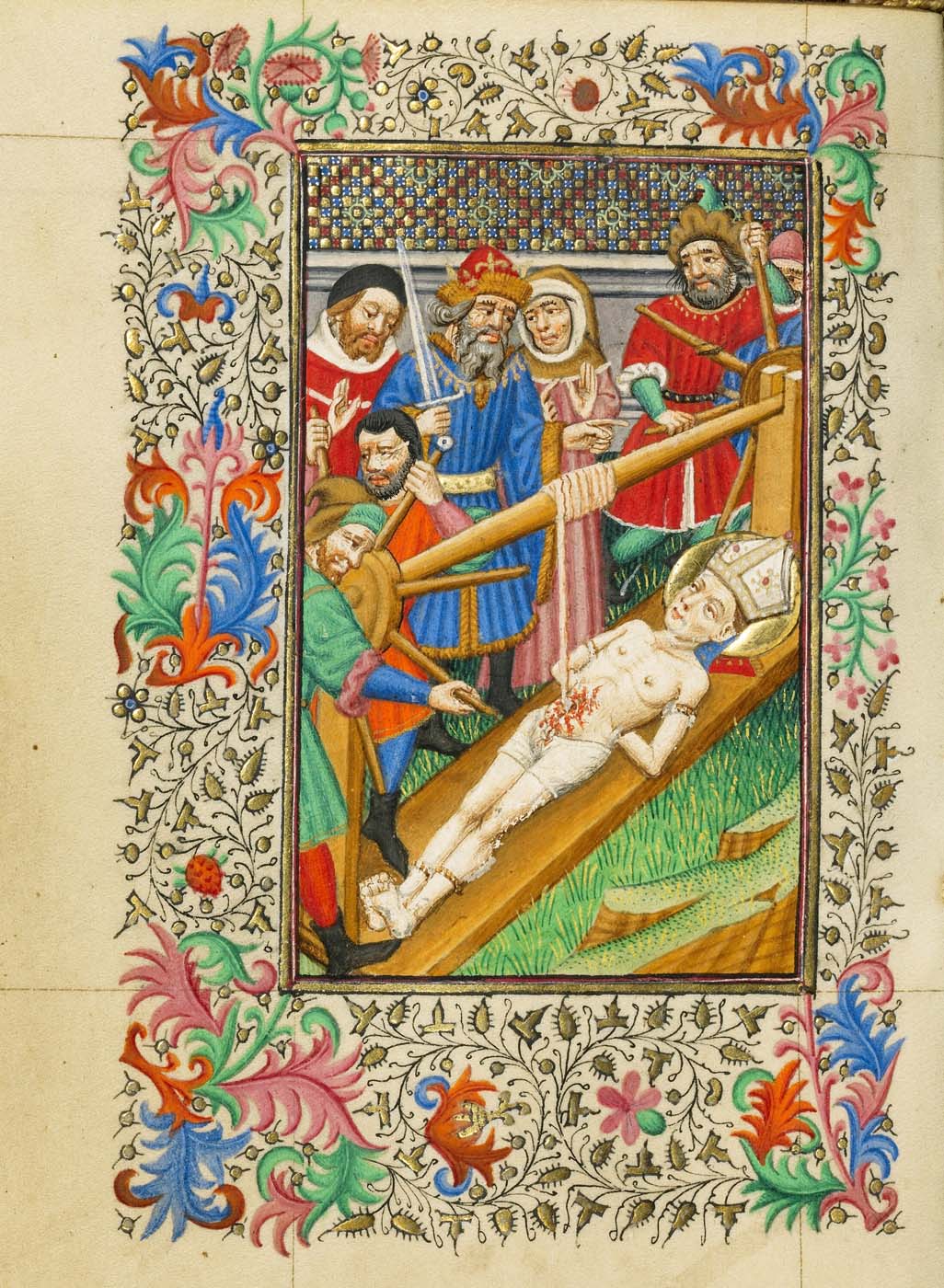
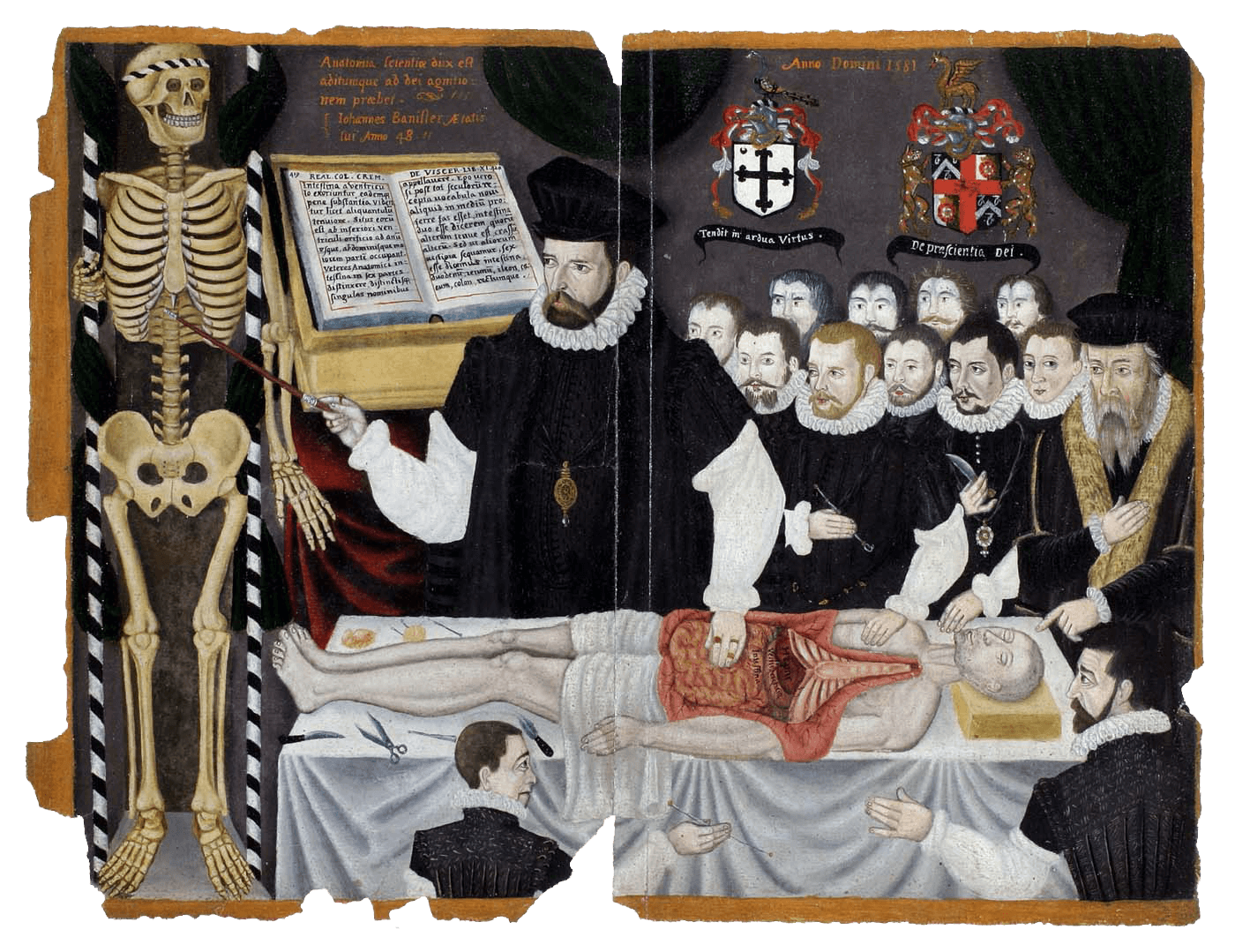
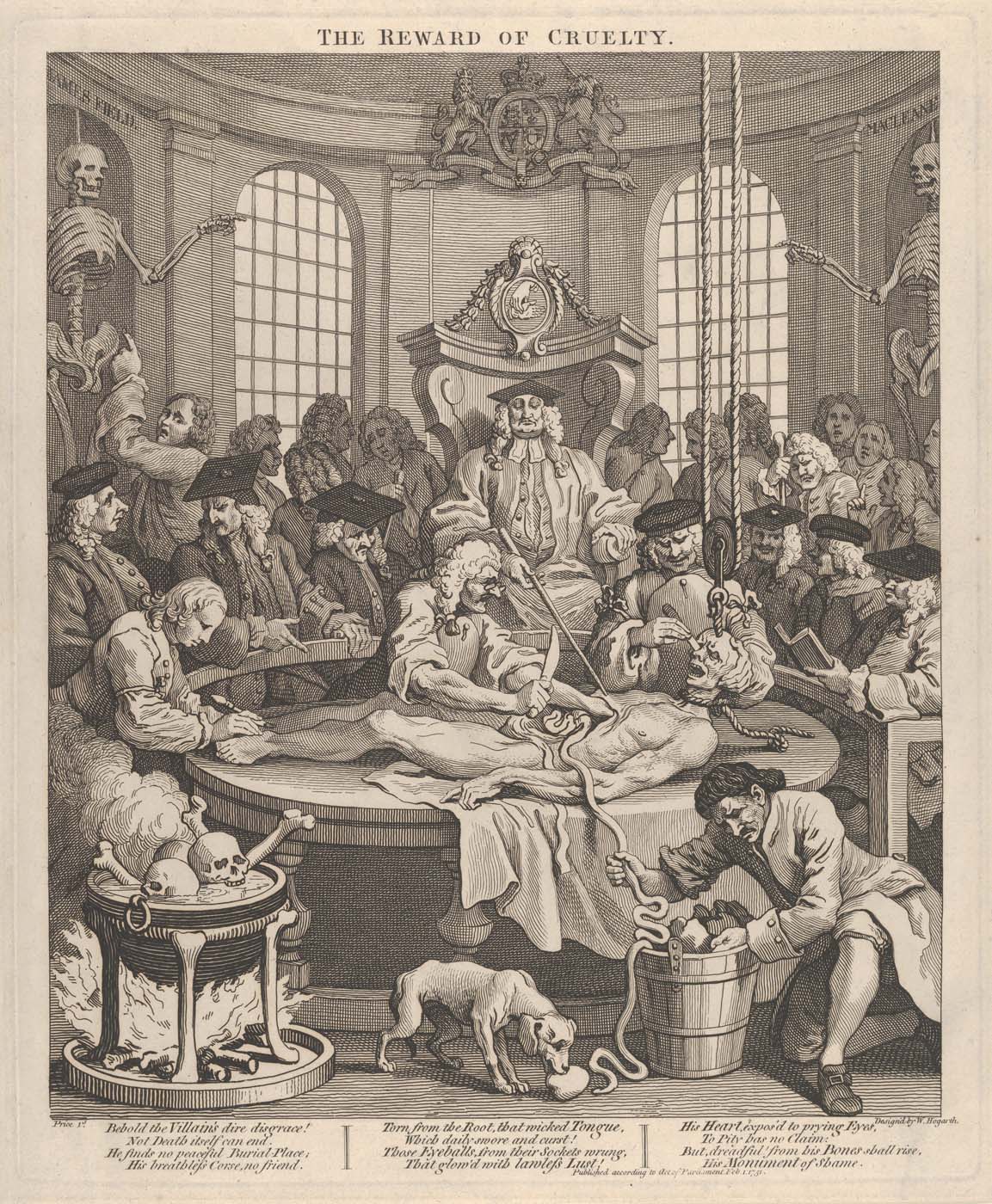
Ranging from the fifteenth to the nineteenth centuries, these images of the viscera were produced for a wide range of purposes: for religious contemplation and for social commentary, as explanatory illustrations of anatomy and of pathology, and as records of anatomical instruction. While reciting a prayer for the safety of their “body and soul”, the owner of a fifteenth-century decorated prayer book could contemplate the equanimity with which St Erasmus endured the removal of his intestines (fig. 1), a sign of the power of his faith in God. The great length of the intestines is indicated as they are wound around a windlass above the saint, and in fig. 4, another scene of disembowelment. In this period in Europe, human dissection was a standard element of medical education, with lectures delivered in front of the open body (figs 34), usually that of an executed criminal. The intestines and stomach were typically the first parts to be removed, as these were the soonest to putrefy. In dissections, knowledge of the body was gained by sight and by touch and, often, by the reading of anatomy texts, like that seen open in the background of John Banister’s late sixteenth-century lecture on the viscera (fig. 3). The painting commemorates the lecture on the viscera given to the Company of Barbers Surgeons in London and was once part of a manuscript with depictions of anatomy and of surgical instruments. 
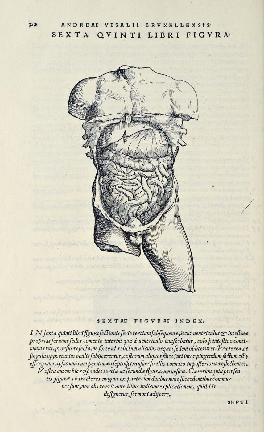
The woodcut from Vesalius’s 
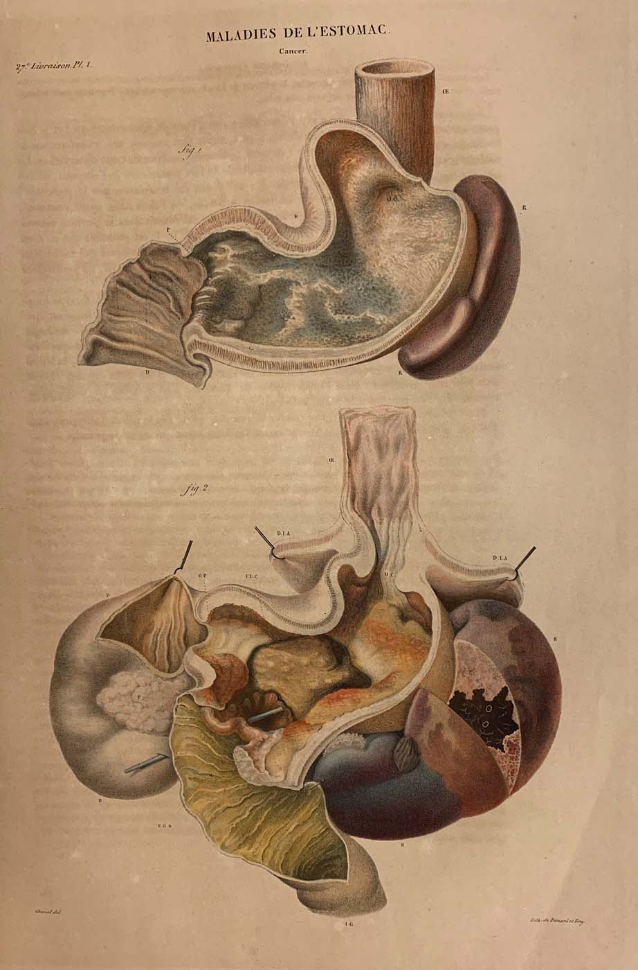
De fabrica (fig. 2) is one in a sequence in which the abdomen is gradually dissected on the page. All the parts are identified in an explanatory text, keyed to the lettering, and the image is also referenced in the main text. Three centuries later, the use of colour printing is used to its full advantage in Cruveilhier’s Anatomie pathologique  (fig. 5) to capture the ravages of cancer in pathological images of the stomach which serve as illustrations linked to case histories, including specific clinical signs and patient’s symptoms.