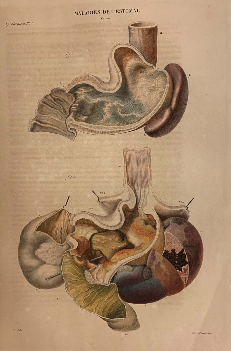
lithograph printed by Benard and Frey after Antoine Touissant de Chazal (French, 1793-1854). From Jean Cruveilhier, Anatomie pathologique du corps humain (Paris: J. B. Baillière, 1829-1842), vol. 2, part 27, pl. 1.
Huntington Library, Art Museum and Gardens, San Marino, California, RB 632011.
–
By the nineteenth century, publishing technology allowed for the introduction of highly detailed colored images. Cruveilhier’s two-volume Anatomie pathologique (1829-35), which initially appeared in short installments, was, at the time, the most comprehensive color-illustrated treatise of the diseases of the human body accounting for both diseased organs and body parts. In a chapter dedicated to diseases of the stomach, these illustrations depicting a type of stomach cancer show changes to the gastric muscle and mucosa. By placing Figure 1 and Figure 2 together on the page, Cruveilhier was able to make comparisons between the organs in his accompanying text. Relying on color—e.g., red and white in the muscular tunic of the stomach in Figure 1—Cruveilhier also indicated differences between healthy and diseased organs, like those in the illustrations, which he associated with particular patients and their histories.