Ca. 1660s-1681c. Lat. Fol. 95, unknown artist
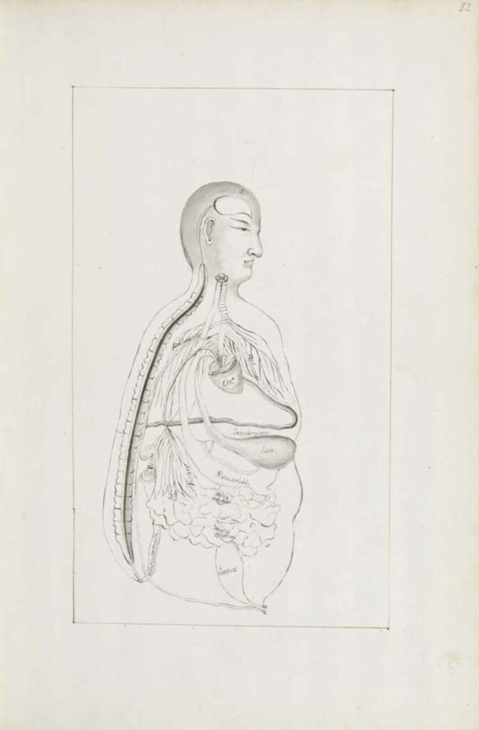
Ca. 1660s-1681c. Lat. Fol. 95, unknown artist This sketch of the “Viscera Man” was also based on a Chinese source (image 1), however, the artist remains anonymous. It was included in the only known extant manuscript (Ms. Lat. Fol. 95 preserved in the Staatsbibliothek in Berlin) of the 1682 printed Specimen Medicinae Sinicae. It illustrates […]
Ca. 1681 Ms sin. 11, part 3 by Christian Mentzel
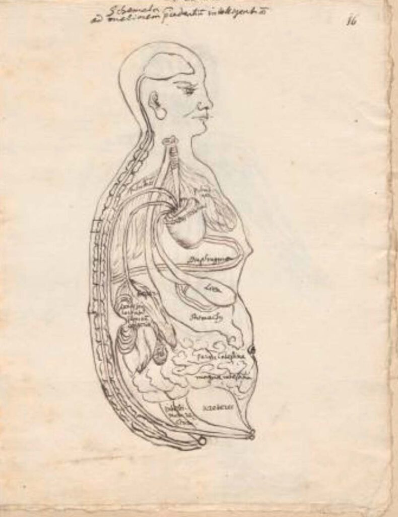
Ca. 1681 Ms sin. 11, part 3 by Christian Mentzel The personal physician to the Great Elector Friedrich Wilhelm of Brandenberg and curator of his library, Christian Mentzel (1622-1701), sketched this “Viscera Man” based on a Chinese source (Image 1). Both Mentzel’s sketch (Image 3) and the printed 1597 original (Image 1) are included in […]
1597 reprint of Wanbing huichun “Diagram of Man’s Side-Body”
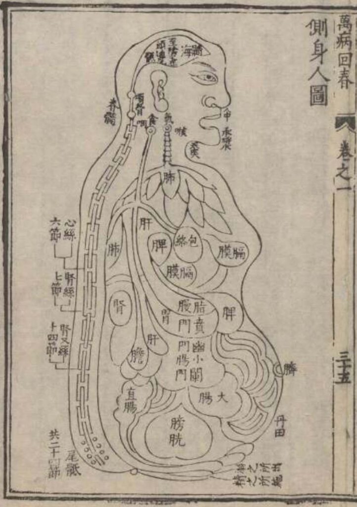
‘Diagram of Man’s Side-Body’ in 1597 reprint of Wanbing huichun (Ceshen ren tu 側身人圖). This version of the “Viscera Man” was printed in a 1597 reprint of the Chinese medical text Myriad diseases ‘Spring Returned’ (i.e., ‘cured’) (Wanbing huichun 萬病回春) that is preserved in the Staatsbibliothek in Berlin. – This 1597 reprint of a “viscera […]
Published in 1597 reprint of a 1565 Yixue gangmu, preserved in Ms sin. 11, part 1, “Enlightened-Hall Diagram of the Viscera”
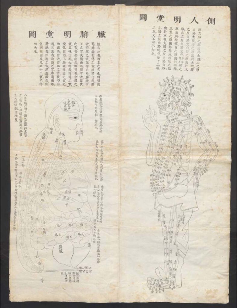
‘Enlightened-Hall Diagram of the Viscera’, in 1597 reprint of a 1565 Yixue gangmu, preserved in Ms sin. 11, part 1 (Zangfu Mingtang tu 臟腑明堂圖). This fold-out plate (ca. 79cm x 58cm) of a “Viscera Man” has been inserted into a manuscript (ms. sin. 11) preserved in Biblioteka Jagiellońska Kraków, Poland, digitalized by the Staatsbibliothek in […]
Diseases of the Stomach (cancer)
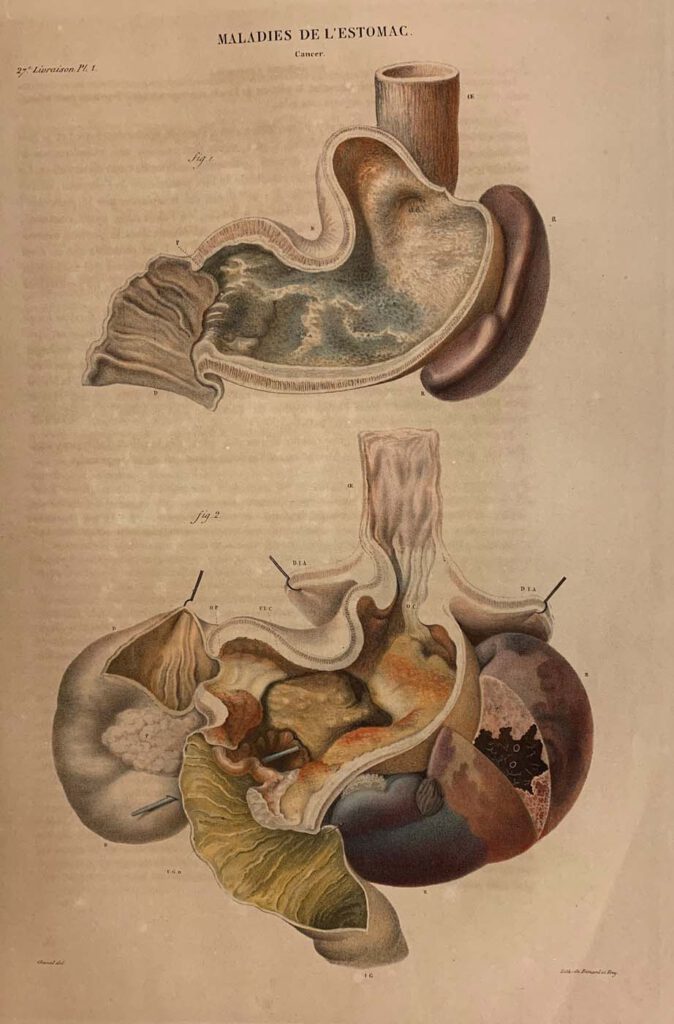
Diseases of the Stomach (cancer) lithograph printed by Benard and Frey after Antoine Touissant de Chazal (French, 1793-1854). From Jean Cruveilhier, Anatomie pathologique du corps humain (Paris: J. B. Baillière, 1829-1842), vol. 2, part 27, pl. 1. Huntington Library, Art Museum and Gardens, San Marino, California, RB 632011. – By the nineteenth century, publishing technology […]
The Reward of Cruelty (The Four Stages of Cruelty)
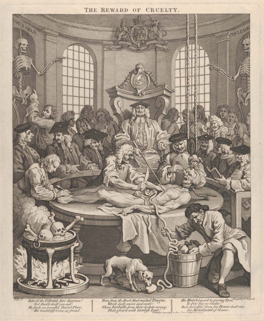
The Reward of Cruelty (The Four Stages of Cruelty), etching and engraving by William Hogarth (English, 1697–1764),February 1, 1751, plate: 15 1/4 x 12 5/8 in. (38.8 x 32 cm)sheet: 15 3/4 x 13 1/16 in. (40 x 33.2 cm) Metropolitan Museum, New York, Gift of Sarah Lazarus, 1891, no. 91.1.139. – This scene, incorporating […]
John Banister Delivering an Anatomical Lecture on the Viscera
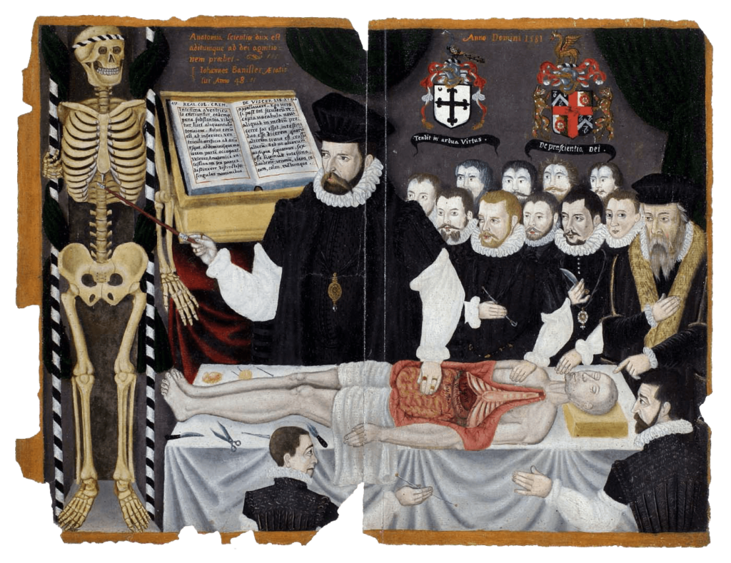
John Banister Delivering an Anatomical Lecture on the Viscera 1581, painting, 33.8 x 43.2 cm. University of Glasgow Library, Glasgow, GB 247 MS Hunter 364 (V.1.1), frontispieceUniversity of Glasgow Archives & Special Collections, MS Hunter 364. – The English surgeon and physician John Banister (1532/3–1599?) lectures on the viscera, placing his hand on the open abdomen […]
Abdominal Dissection
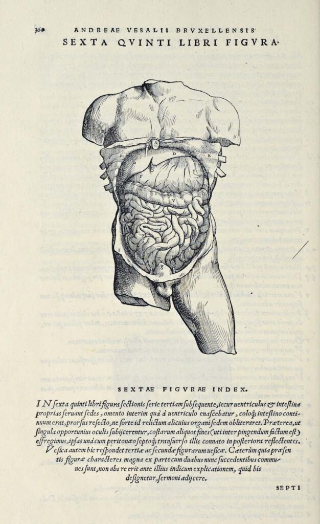
Abdominal Dissection woodcut after Jan Steven van Calcar (North Netherlandish, ca. 1515– ca. 1546). From Andreas Vesalius, De humani corporis fabrica libri septem (Basel: J. Oporinus, 1543), bk. 5, p. 360 [460], fig. 6. Getty Research Institute, Los Angeles, 84-B27611 – Ribs have been broken and the skin peeled back to display the liver, stomach, […]
The Martyrdom of St Erasmus
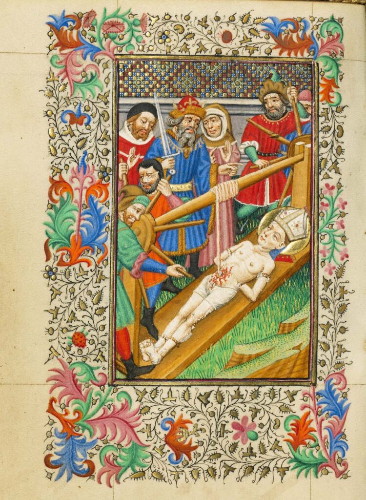
The Martyrdom of St Erasmus about 1430–1440, by Master of Sir John Fastolf (French, active before about 1420 – about 1450). Tempera colors, gold leaf, and ink, Leaf: 12.1 × 9.2 cm (4 3/4 × 3 5/8 in.) The J. Paul Getty Museum, Los Angeles, Ms. 5 (84.ML.723), fol. 38v – This scene of the martyrdom […]
Roman marble version of a dying Gaul
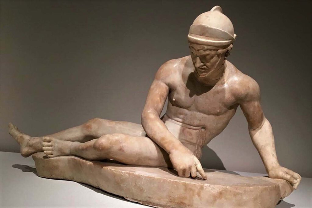
Roman marble version of a dying Gaul the original was part of a major sculptural display on the Athenian Acropolis of Attalid Greeks fighting Gauls in Asia Minor, Gods fighting Giants, Greeks fighting Amazons, and Greeks fighting Persians in the Persian Wars, c.200 B.C. Naples Dying GaulPhoto: Mary Harrsch, CC-BY-NC-SA-2.0 – In c. 200 BCE Attalos […]