Comparative Guts

Search
Close this search box.

Diseases of the Stomach (cancer)

Erkrankungen des Magens (Krebs) Auf der Seite aus Cruveilhiers Anatomie sind unter der Überschrift „Die Krankheiten des Körpers“ zwei farbige Abbildungen des Magens platziert. Sie stellen ein gesundes und ein erkranktes Organ dar. In der oberen Hälfte des Blattes wird ein Magen im Querschnitt gezeigt. Er ist in Form eines liegenden Ovals mit Einbuchtungen und Auswölbungen an den Rändern dargestellt. Die Ränder sind beige. Oben ist der Ansatz der Speiseröhre sichtbar, unten links der des Darms. Das Innere des Magens ist zu zwei Dritteln in Grau dargestellt, das obere Drittel in Beige. Die zweite Abbildung unten auf der Seite zeigt den Magen im Querschnitt mit den Ansätzen von Speiseröhre und Darm wie oben beschrieben. Rechts daneben ist ein Organ in braun abgebildet. Es hat eine ebenfalls ovale Form, in der ein keilförmiger Ausschnitt erkennbar ist. Darin befindet sich ein schwarzer Fleck mit gezackten Rändern. Das Innere des Magens ist in dieser Darstellung in verschiedenfarbige Bereiche unterteilt. In der Mitte liegt eine ockerfarbene nahezu kreisförmige Fläche mit einem schwarzen Umriss. Sie füllt fast den gesamten Innenraum aus. Die wulstige Fläche rechts daneben ist in Rottönen dargestellt, eine dunkelrote trichterförmige Fläche markiert den Übergang zum ockerfarbenen Darm. John Baptiste Cruveilhier war ein französischer Arzt, Anatom und Pathologe, der im 19. Jahrhundert in Limoges, Nordwestfrankreich, praktizierte. Seine Erkenntnisse schrieb er unter anderem in seiner Anatomie Pathologique nieder und schuf damit ein fundamentales Werk. — — Diseases of the Stomach (Cancer) On the page from Cruveilhier's Anatomy, two coloured illustrations of the stomach are placed under the heading “The Diseases of the Body.” Two organs are depicted, one healthy and one diseased. In the upper half of the page, a stomach is shown in cross-section. It is depicted in the form of a reclining oval with indentations and bulges at the edges. The edges are beige. At the top, the base of the oesophagus is visible, and that of the intestine is at the bottom left. Two-thirds of the inside of the stomach is shown in grey, and the upper third in beige. The second illustration at the bottom of the page shows the stomach in cross-section with the attachments of the oesophagus and intestine as previously described. To the right, an organ is shown in brown. It also has an oval shape in which a wedge-shaped cutout can be seen. Within it is a black spot with jagged edges. In this illustration, the inside of the stomach is divided into different coloured areas. In the middle is an ochre-coloured almost circular area with a black outline. It fills almost the entire interior. The bulging area to the right is depicted in shades of red, and a dark red funnel-shaped area marks the transition to the ochre-coloured intestine. John Baptiste Cruveilhier was a French physician, anatomist and pathologist who practised in Limoges, northwestern France, during the 19th century. Among other things, he wrote down his findings in his "Anatomie Pathologique" and thus created a fundamental work.

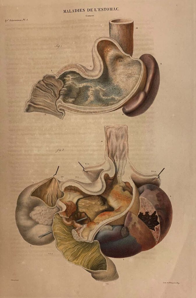
Diseases of the Stomach (cancer) lithograph printed by Benard and Frey after Antoine Touissant de Chazal (French, 1793-1854). From Jean Cruveilhier, Anatomie pathologique du corps humain (Paris: J. B. Baillière, 1829-1842), vol. 2, part 27, pl. 1.  Huntington Library, Art Museum and Gardens, San Marino, California, RB 632011. – By the nineteenth century, publishing technology […]

The Reward of Cruelty (The Four Stages of Cruelty)

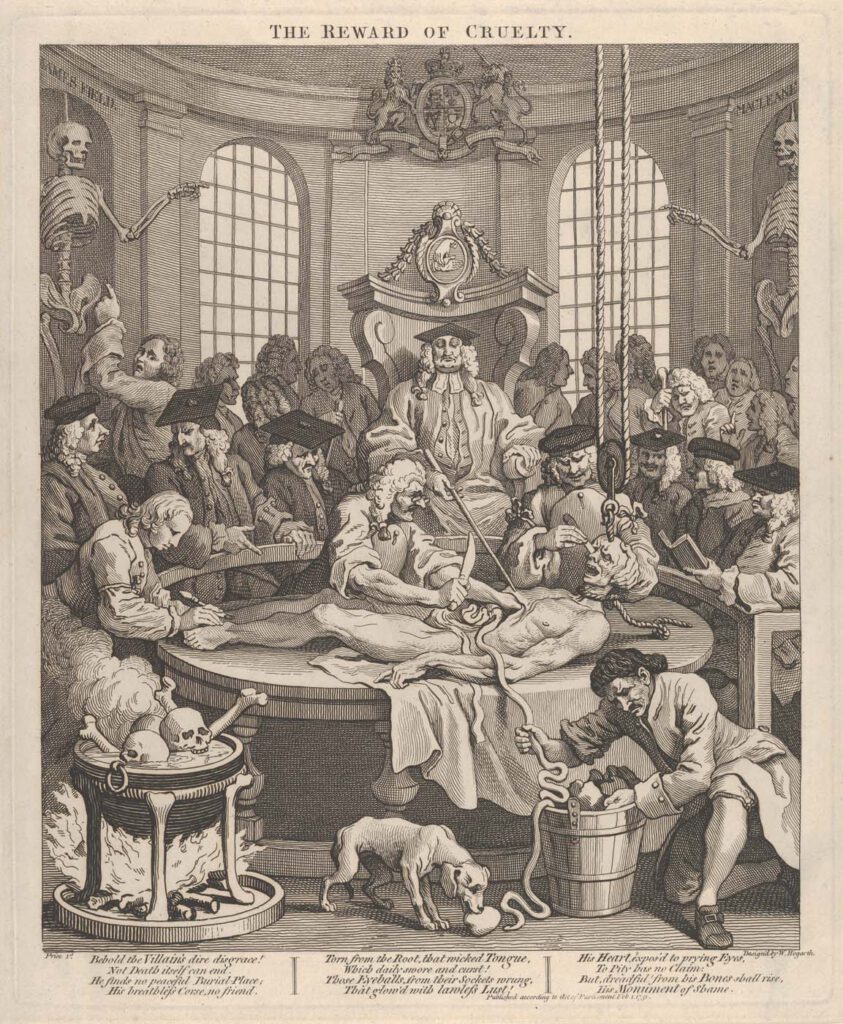
The Reward of Cruelty (The Four Stages of Cruelty), etching and engraving by William Hogarth (English, 1697–1764),February 1, 1751, plate: 15 1/4 x 12 5/8 in. (38.8 x 32 cm)sheet: 15 3/4 x 13 1/16 in. (40 x 33.2 cm) Metropolitan Museum, New York, Gift of Sarah Lazarus, 1891, no. 91.1.139. – This scene, incorporating […]

John Banister Delivering an Anatomical Lecture on the Viscera

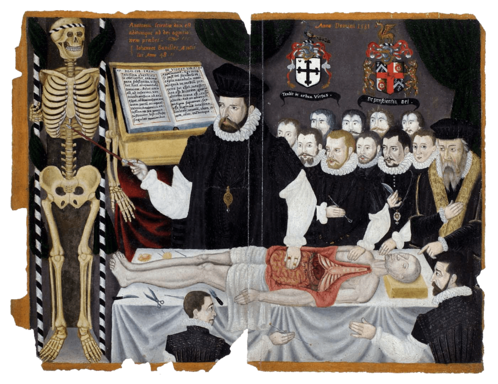
John Banister Delivering an Anatomical Lecture on the Viscera 1581, painting, 33.8 x 43.2 cm. University of Glasgow Library, Glasgow, GB 247 MS Hunter 364 (V.1.1), frontispieceUniversity of Glasgow Archives & Special Collections, MS Hunter 364. – The English surgeon and physician John Banister (1532/3–1599?) lectures on the viscera, placing his hand on the open abdomen […]

Abdominal Dissection

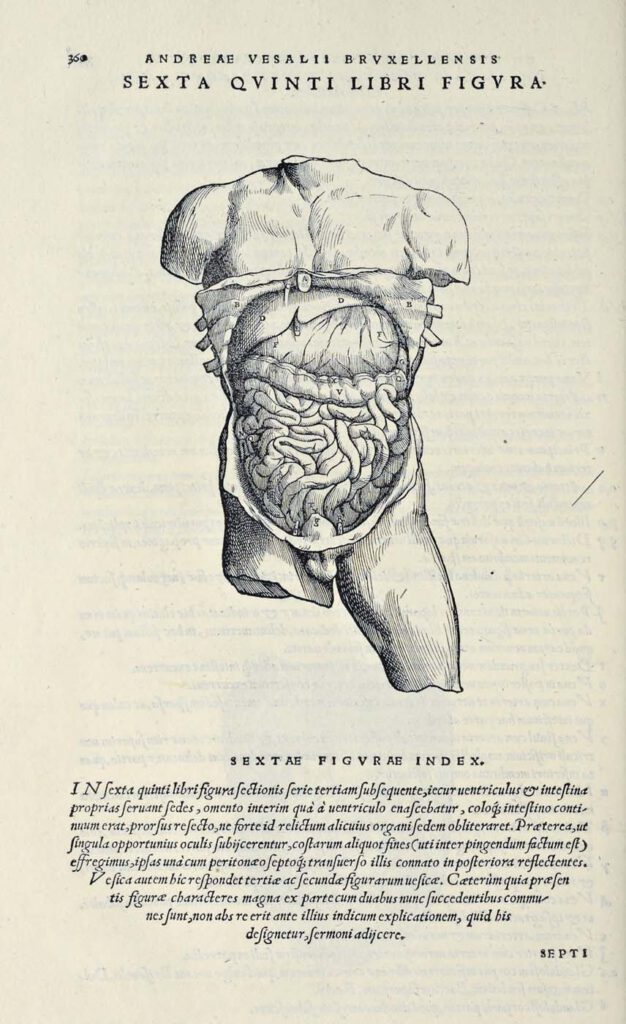
Abdominal Dissection woodcut after Jan Steven van Calcar (North Netherlandish, ca. 1515– ca. 1546). From Andreas Vesalius, De humani corporis fabrica libri septem (Basel: J. Oporinus, 1543), bk. 5, p. 360 [460], fig. 6.  Getty Research Institute, Los Angeles, 84-B27611 – Ribs have been broken and the skin peeled back to display the liver, stomach, […]

The Martyrdom of St Erasmus

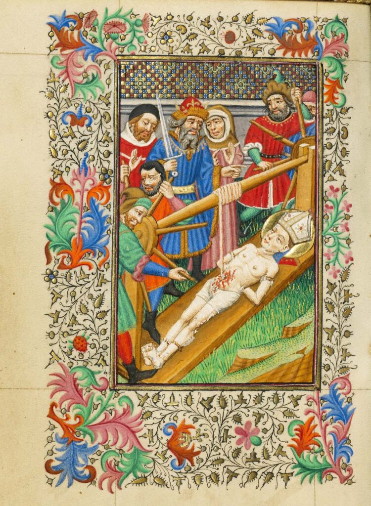
The Martyrdom of St Erasmus about 1430–1440, by Master of Sir John Fastolf (French, active before about 1420 – about 1450). Tempera colors, gold leaf, and ink, Leaf: 12.1 × 9.2 cm (4 3/4 × 3 5/8 in.) The J. Paul Getty Museum, Los Angeles, Ms. 5 (84.ML.723), fol. 38v – This scene of the martyrdom […]