Henry Gray (anatomist) and Henry Vandyke Carter (artist), Fig. 1005, Superior and inferior duodenal fossæ.
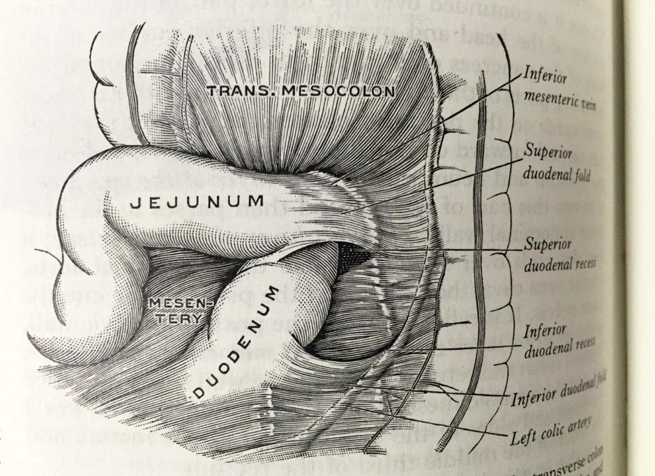
Henry Gray (anatomist) and Henry Vandyke Carter (artist), Fig. 1005, Superior and inferior duodenal fossæ. Gray’s Anatomy Descriptive and Applied. A New American Edition (1913), p. 1265.Private collection: Nina Sellars. Photographer: Nina Sellars. – Giving thoughtful attention to the physical qualities of the classical anatomical atlases brings our awareness not only to their content and […]
John Sloan, A self-portrait of the artist holding a cup of barium and undergoing an upper gastrointestinal fluoroscopic study under the care of two radiologists, 1926.
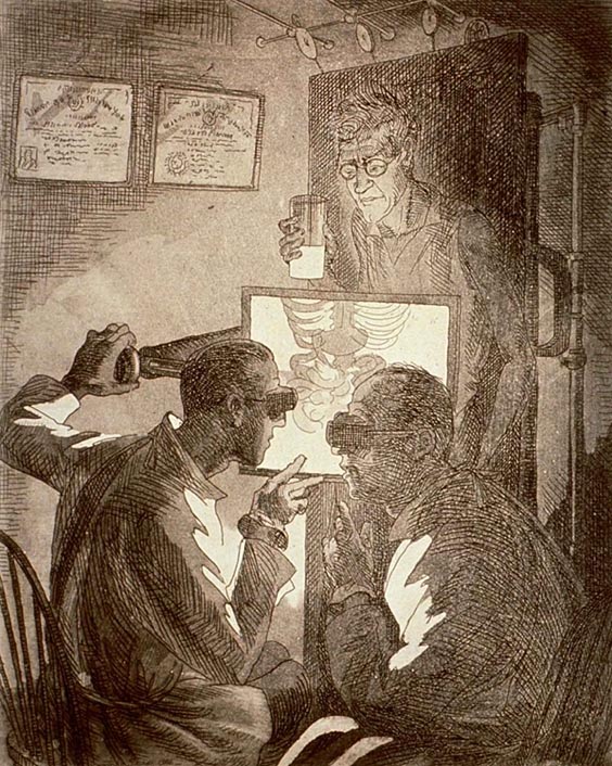
John Sloan, A self-portrait of the artist holding a cup of barium and undergoing an upper gastrointestinal fluoroscopic study under the care of two radiologists, 1926. Medium: etching and aquatint. Collection: National Library of Medicine. – Anatomy is a science predominantly based on visual observation. Therefore, the anatomical body’s existence is seemingly reliant on modes […]
Bernardino Capitelli, Anatomical Dissection, c. 1604 – 1639.
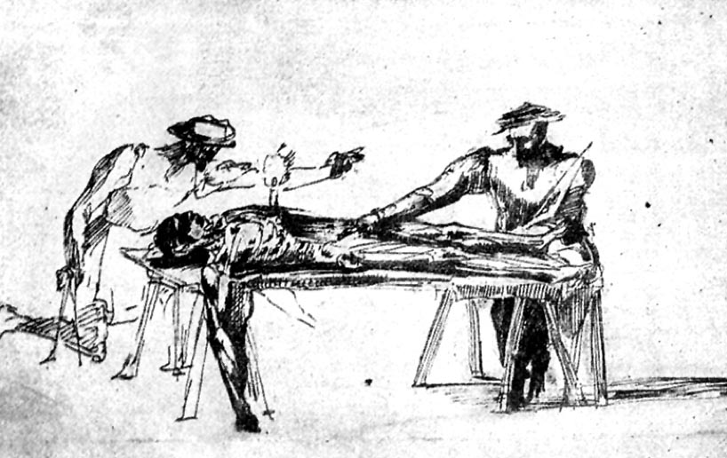
Anatomical Dissection, 1495-approximately 1543 Two men studying a corpse by the light of a candle stuck in itschest. Etching after a drawing attributed to Polidoro Caldara (Polidoro da Caravaggio). Wellcome Collection. Attribution 4.0 International (CC BY 4.0) – In the Renaissance era, the tempo and sequence of a dissection were dictated by the decomposition rate […]
Andreas Vesalius (anatomist) and Jan van Calcar (artist), studio of Titian. The Seventh Plate of Muscles.
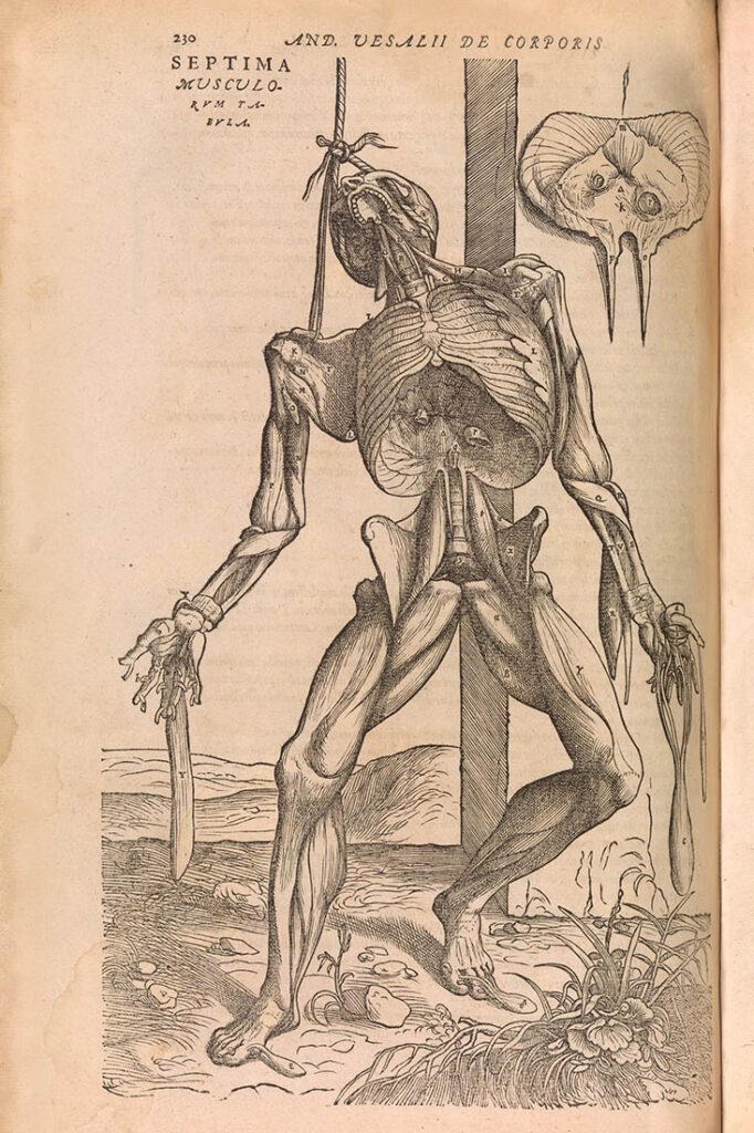
Andreas Vesalius (anatomist) and Jan van Calcar (artist), studio of Titian. The Seventh Plate of Muscles. Illustration from the second edition of the anatomy text De humani corporis fabrica (On the Fabric of the Human Body), 1555, p. 230. Medium: woodblock print made by unknown block cutter. Metropolitan Museum of Art, New York, Gift of […]
Dancer Shona Dunlop performing the role of Cain in Cain and Abel, Sydney, Australia, 1940.
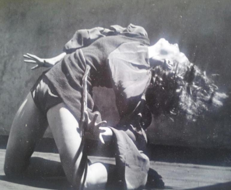
Dancer Shona Dunlop performing the role of Cain in Cain and Abel, Sydney, Australia, 1940. Choreographer: Gertrud Bodenwieser. Composer: Marcel Lorber. Medium: gelatin silver photograph. Photographer: Margaret Michaelis. Collection: Hocken Library Collection at University of Auckland. – Shona Dunlop was born in Ōtepoti, Aotearoa (Dunedin, New Zealand), and studied with choreographer and dancer Gertrud Bodenwieser […]