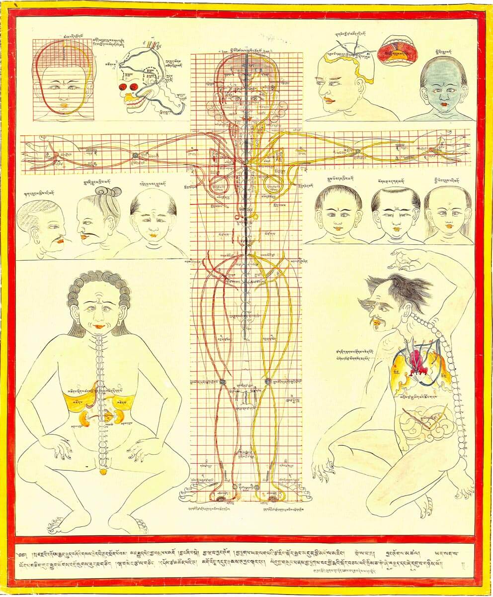
Major connecting blood vasculature of body including gut with related vulnerable points
20th century reproduction of 17th century CE painting. Held in Ulan-Ude, Republic of Buryatia, Russia.
–
Explicated in high detail in the traumatology section of the Four Tantras as developed from early military medical manuals, this painting depicts major blood vessels of the upper and lower abdomen, head, neck, and extremities. Major wound types are also noted. Blood vessels of the head and aortic path adjacent to the spine are shown in azure color, those of the neck in arsenic yellow, those of the trunk in vermillion, and extremities in black with Tibetan alphabetical symbols. Vessels on the figure’s right side are in red and left side in yellow; capillary beds demonstrating arterial-venal transition are grey circular swirls. Blood vessels specific to the gut and other vessel organs are illustrated on both sides. Internal blood vessels perfusing blood to small and large intestine are diagrammed specifically. Radial artery for pulse analysis is labeled “observation vessel” (blta rtsa) and ulnar for assessing longevity is la vessel (bla rtsa).