Comparative Guts

Search
Close this search box.

1300–1800 CE

Arabic and Persian Materials

Ignacio Sanchez

Senior Research Fellow Classics and Ancient History, University of Warwick
Introduction
The terms Islamic or Arabic medicine are, as often noted, misleading. Many of the physicians who lived and worked in pre-modern Islamic societies were neither Muslim nor Arab, and the shortcomings of defining multi-ethnic and culturally varied regions in religious terms are obvious, let alone its ideological implications (1). Yet, the use of anatomical representations in the medical traditions developed in the Islamic world is determined, to a great extent, by religious concerns. The Arabic term for anatomy –later adopted in Persian and Ottoman Turkish– is tashrīḥ, which literally means “dissecting”. Ironically, the medieval sources contain very little references to human dissection, since this practice was regarded as contrary to religious principles. (2) The anatomical paradigm to which these physicians adhered was Galenic and the opinions of Galen were rarely questioned. As Maimonides said when discussing Galen’s works on anatomy, no physician had practiced dissection after him. To this limitation, with obvious consequences for the knowledge of the internal organs, we should add yet another religious constrain: the prohibition of human representation, defended by a relevant number of Muslims scholars. As a consequence, anatomical representations in pre-modern Islamic societies are rare. Most of them are connected to a single pictorial tradition inaugurated by Manṣūr ibn Ilyās in the 14th century and were used in medical works that were produced and circulated in the Eastern Mediterranean and Middle Eastern Islamic lands. (1) On this tradition see the introduction to Peter Pormann and Emilie Savage-Smith (2007) (2) For a discussion on this topic see Emilie Savage-Smith (1995).
Guts and arabic materials
The first example of anatomical illustrations in an Islamic milieu was a Persian translation from Chinese, the Tansūqnāma, commissioned by the Mongol vizier Rashīd al-Dīn in 1313 (figs. 1 and 2). A second anatomical work written in Persian by Manṣūr ibn Ilyās, which included full-page representations of the human body, was also composed in 1386 for a Timurid ruler (figs. 3 and 4). Prior to the 14th century we only find a few schematic representations of the eyes and the brain ventricles that use geometric forms. (1) The Tansūqnāma has survived in a unique manuscript and did not seem to have been copied or originated any textual or pictorial tradition. In contrast, the second of the aforementioned works, the so-called 
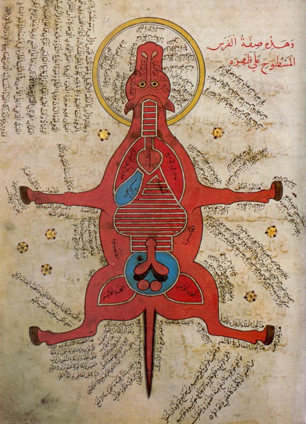
al-Tashrīḥ al-Manṣūrī, became a model for later representations of both human and animal bodies, not only in the Islamic Mediterranean and Middle Eastern lands, but also South Asia and the Tibet. This tradition is known as the 
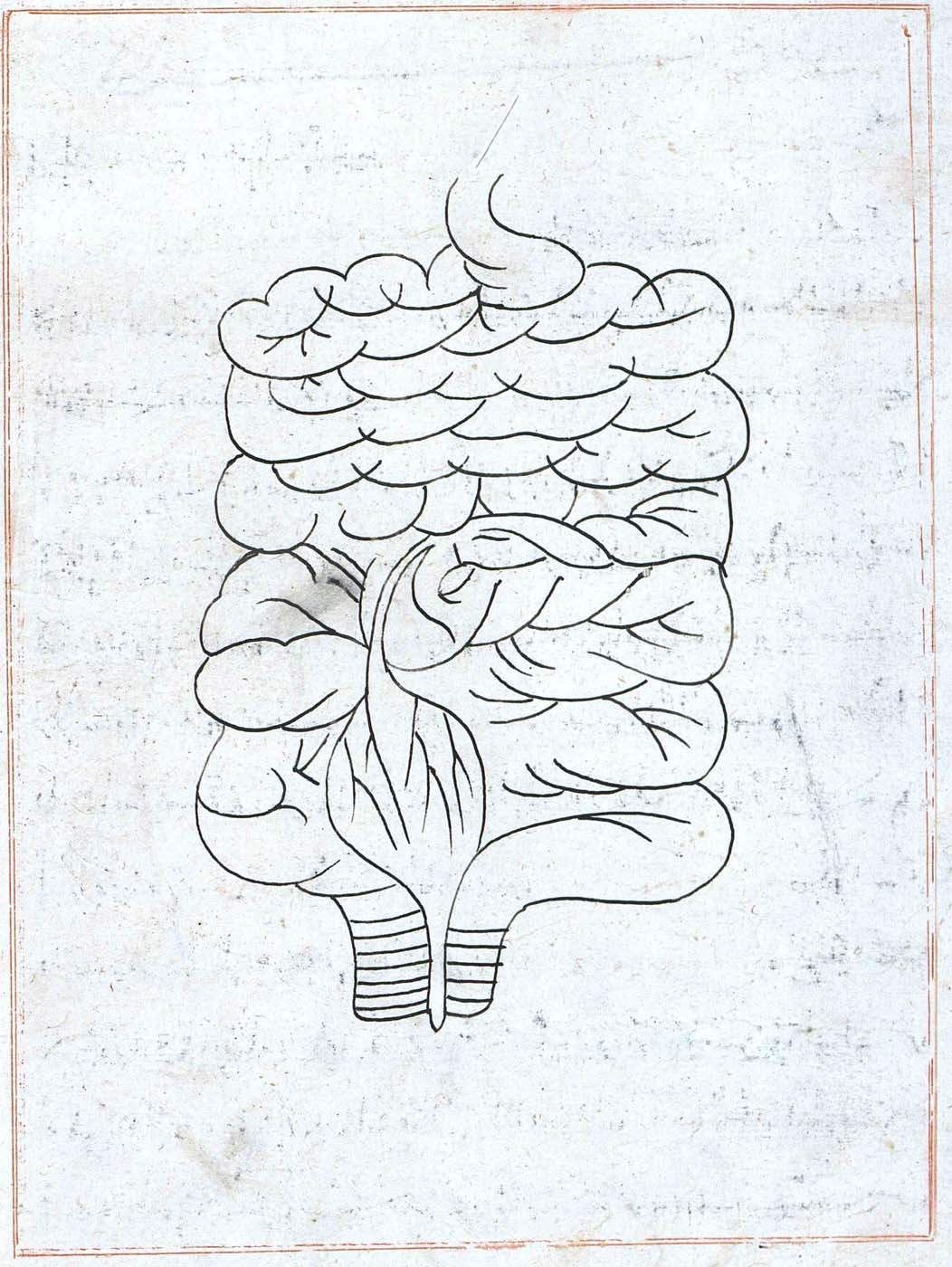
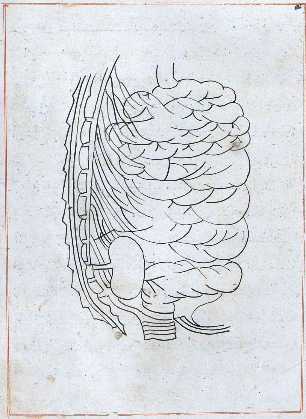
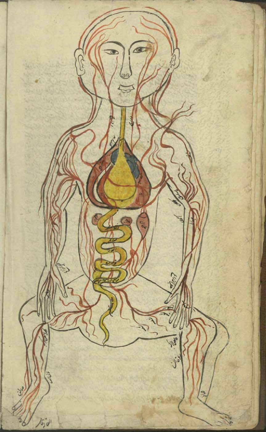
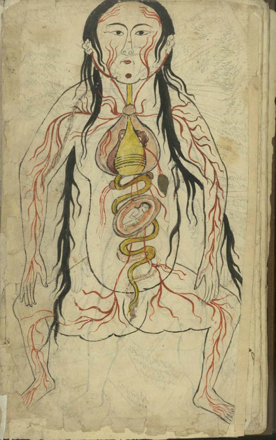
Fünfbilderserie (‘the Five-images series’), since it usually includes five illustrations representing the silhouette of a “flat man”, as seen from above, and depicting arteries, veins, nerves, bones, and muscles respectively. Some sets of illustration sometimes include a representation of a pregnant woman with the foetus in the womb (fig. 4). The origin of these illustrations has been traced back to 12th century Europe (2) In addition to the exceptional illustrations taken from Chinese works in the Tansūqnāma, the only representations of the internal organs that have come down to us from pre-modern Islamic lands are those belonging to the Fünfbilderserie tradition. The success of this pictorial model went beyond the works on human anatomy and similar designs were later used in the illustration of veterinary works (fig. 5). Neither the Tansūqnāma  nor al-Tashrīḥ al-Manṣūrī have been edited or properly studied. The use of these visual representations and their relationship with the anatomical or veterinary texts they accompany also awaits a proper research. The depiction of the internal organs in the figures copied from Chinese sources in the Tansūqnāma is realistic and their position within the body roughly accurate. In the Fünfbilderserie tradition, the depiction of the anatomical parts is rather schematic, although their position is in general accurate. The organs that participate in the digestion of food according to the Galenic medical tradition are often drawn in the same colour. In general, the guts are not of special symbolic or metaphorical significance in the cultural traditions conveyed by these texts, in contrast with other organs such as the brain, the heart and the liver. (3) See Emilie Savage (2007). (4) See Ynez Violé O’Neill (1977), Andrew Newman (1998).
References

Peter Pormann and Emilie Savage-Smith. 2007. Islamic Medicine. Edinburgh: Edinburgh University Press.

Emilie Savage-Smith, “Attitudes towards Dissection in Medieval Islam,” Journal of the History of Medicine and Allied Sciences 50/1 (1995): 67-110.

Emilie Savage-Smith. 2007. “Anatomical Illustrations in Arabic Manuscripts”, in Anna Contadini (ed.), Arab Painting: Text and Image in Illustrated Arabic Manuscripts (Leiden and Boston: Brill) 147-159.

Ynez Violé O’Neill. 1977. “The Fünfbilderserie – A Bridge to the Unknown” in Bulletin of the History of Medicine 51 (1977): 538-549

Andrew Newman. 1998. “Tašrīḥ-i Manṣūrī: Human Anatomy between the Galenic and Prophetical Medical Traditions” in Ž. Vesel, H. Beikbaghban, and B. Thierry de Crussol des Epesse (eds.), La science dans le Monde Iranien à l’époque islamique (Tehran: Institut Français de Recherche en Iran): 253-272.