Illustration accompanying De Arte Physicali et de Cirurgia
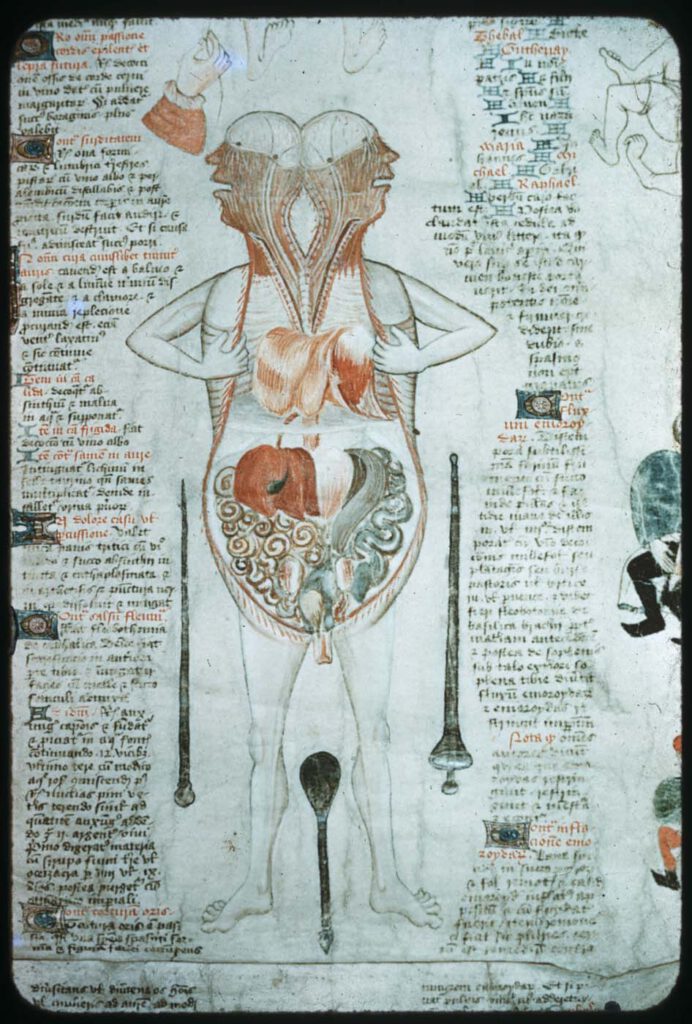
Illustration accompanying De Arte Physicali et de Cirurgia, attributed to John Arderne Stockholm, Kungliga Biblioteket X.118, fol. 6v/6r; c. 1430 – While the frontal approach to anatomical illustration exhibited in the Five/Nine-Figure Series and Guido da Vigevano’s work is most dominant, an alternative, sagittal approach emerged in the medieval period and survives in a few […]
Guido da Vigevano’s Anathomia, Figure 8
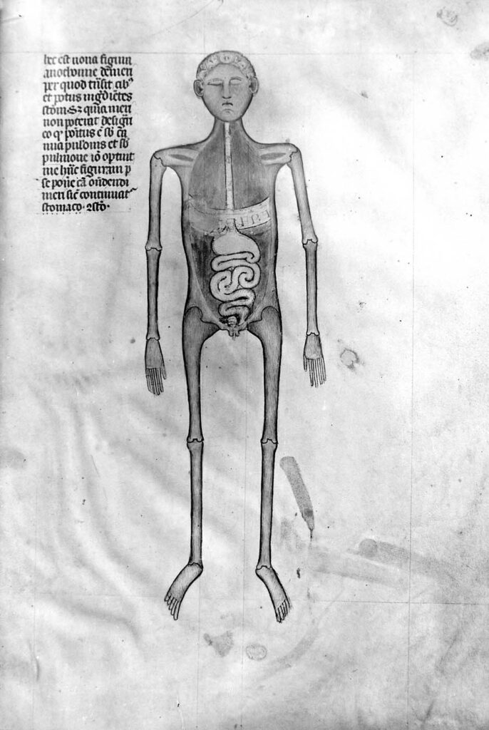
Guido da Vigevano’s Anathomia, Figure 9 (1345) Guido da Vigevano, Anathomia, 1345CC BY-NC-SA 4.0 – Although human dissection began to be systematically practiced in Italy in the early 14th century, it was not immediately embraced across Europe. This prompted Guido da Vigevano to produce a series of seventeen illustrations included in his Book of Notable […]
Organs of the abdomen
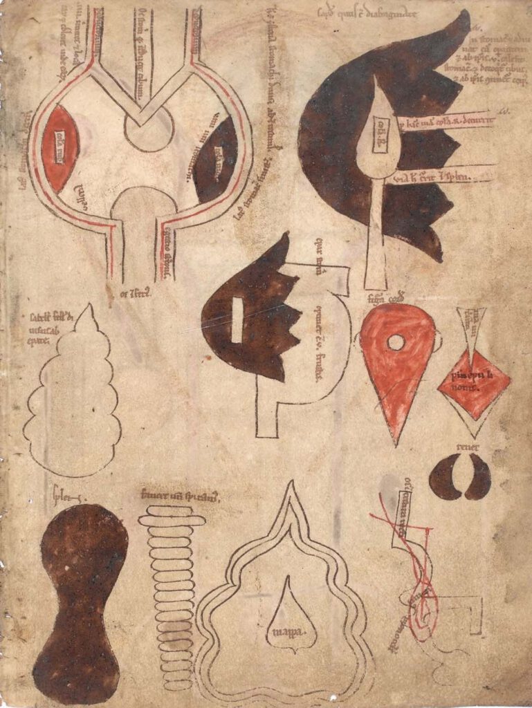
Organs of the abdomen Cambridge, Gonville and Caius College, MS 190/223, fol. 5r; 12th cent. CE Photo: By Permission of the Master and Fellows of Gonville and Caius College, Cambridge. – While the five figures representing the five bodily systems are the core of the Five-Figure Series, several iterations include nine figures rather than five, […]
Anatomical figures
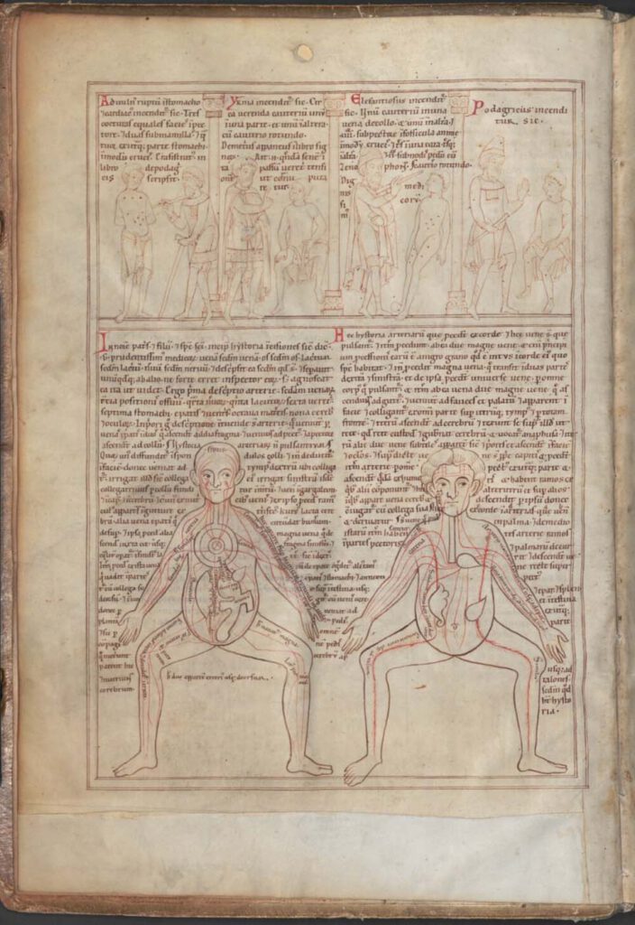
Anatomical figures (Munich, Bayerische Staatsbibliothek, Clm 13002, fol. 2v; 1165 CE) Photo: Bayerische Staatsbibliothek MünchenCC BY-NC-SA 4.0 – These figures offer the earliest example of the so-called Five-Figure Series (Fünfbilderserie), which appears in medical manuscripts from across the medieval world, with Latin, Provençal, Arabic, and Persian versions surviving. The series, which likely originated in Late […]
Illustrations of the uterus
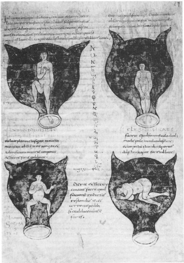
Illustrations of the uterus from Muscio’s epitome of Soranus’ Gynecology Brussels, Bibliothèque Royale MS 3714, fol. 28r; c. 900 CECC0 1.0 Universal (CC0 1.0) – These illustrations of various positions of the fetus in the womb accompany Muscio’s Late Antique Latin summary of Soranus’ 2nd century Greek work on gynecology. The 9th/10th century manuscript pictured […]