Comparative Guts

Search
Close this search box.

Manase Dōsan 曲直瀬道三, Hyakufuku zusetsu
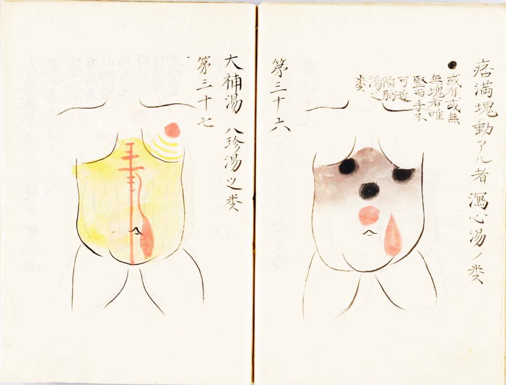
Manase Dōsan 曲直瀬道三, Hyakufuku zusetsu (百腹図説, “Illustrated Explanations of a Hundred Bellies”; n.d.) – Nothing more directly translated the early modern stress on practical, tangible realities than the Japanese innovation known as fukushin 腹診 (abdominal inspection). Whereas Chinese doctors had relied chiefly on feeling the pulse at the wrist to diagnose diseases, Japanese advocates of […]

Sugita Gempaku’s Kaitai shinsho
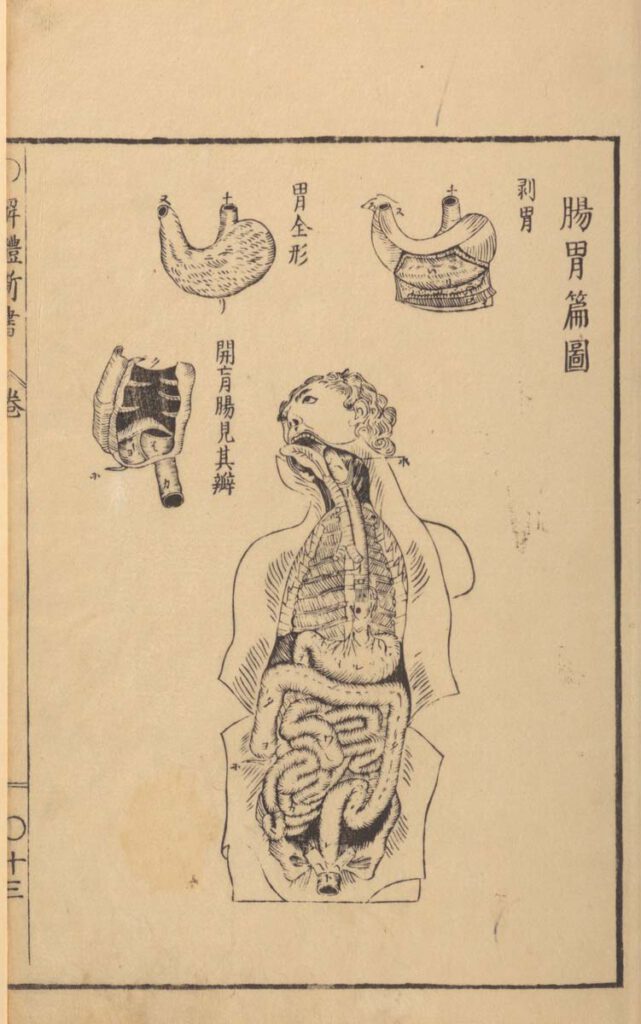
Sugita Gempaku’s Kaitai shinsho "Neues Buch der Anatomie" — Diese hochformatige Schwarz-weiß-Zeichnung ist ein geöffneter Torso mit Kopf abgebildet, darüber und daneben Detailansichten von Organen. Neben jeder Detailansicht verlaufen japanische Schriftzeichen von oben nach unten. Das Original der hochformatigen Zeichnung stammt aus dem 18. Jahrhundert. Der Torso mit Kopf nimmt in der Höhe zwei Drittel der Darstellung ein. Er ist zentral positioniert. Der Kopf mit kurzen lockigen Haaren ist leicht gedreht und nach hinten geneigt. Die Augen sind geöffnet. Der Unterkiefer fehlt, so dass die Zungenunterseite und die obere Zahnreihe sichtbar sind. Aus dem Rachen führen Speise- und Luftröhre in den Brustraum, wo die Rippen angedeutet sind. Die Speiseröhre mündet in ein quer liegendes sackförmiges Organ, den Magen. Nach links tritt aus dem Magen der Dünndarm heraus. Der windet sich in einem Bogen nach oben und anschließend in Schlingen und Schlaufen nach unten. Dieses Knäuel wird eingerahmt vom Dickdarm. Er verläuft unterhalb des Magens nach rechts und dann senkrecht nach unten, wo er nach einer engen Schlaufe in den Mastdarm mündet. Einzeln stehende japanische Schriftzeichen sind an Magen und Darm erkennbar. Die Ränder des geöffneten Brust- und Bauchraumes werden von Gewebestreifen gesäumt, die wie zur Seite geklappt wirken. Das Organ, das links oberhalb des Kopfes vergrößert abgebildet ist, ist ein Magen. Es entspricht in Form und Gestaltung in etwa dem Magen im Torso. Rechts daneben befindet sich ein weiterer Magen, bei dem die Unterseite aufgeklappt ist. Links neben dem Kopf ist der Übergang vom Dünn- in den Dickdarm durch eine kammerartig strukturierte Illustration veranschaulicht. —— Gempaku's Kaitai shinsho “New Book of Anatomy” — This portrait format black and white drawing depicts an open torso with a head, with detailed views of organs above and beside it. Next to each detailed view, Japanese characters run from top to bottom. The original drawing dates from the 18th century. The torso with a head occupies two-thirds of the height of the depiction. It is positioned centrally. The head with short curly hair is slightly turned and tilted backwards. The eyes are open. The lower jaw is missing, so that the underside of the tongue and the upper row of teeth are visible. From the pharynx, the oesophagus and trachea lead into the chest cavity, where the ribs are indicated. The oesophagus opens into a transverse sac-shaped organ, the stomach. The small intestine emerges from the stomach to the left and winds upwards in an arc and then downwards in loops and loops. The small intestine is framed by the large intestine. It runs below the stomach to the right and then vertically downwards where, after a narrow loop, it flows into the rectum. Single-standing Japanese characters are visible on the stomach and intestine. The edges of the opened chest and abdomen are lined with strips of tissue that appear to be folded to the side. An enlarged organ is depicted above the head and on the left, it is the stomach. It roughly corresponds in shape and design to the stomach within the open torso. To the right of it is another enlarged stomach with its underside open. To the left of the head, the transition from small to large intestine is illustrated by a chamber-like structured illustration.

Sugita Gempaku’s Kaitai shinsho (解体新書, “New Book of Anatomy”; 1774) – Sugita Gempaku undertook his epoch-making translation of a Dutch anatomical manual before he could read Dutch. This point is key: What persuaded him of the manual’s truth was not its text, which he initially couldn’t comprehend, but rather, its images, and more specifically their […]

Treatment points for moxibustion, venesection, and surgery
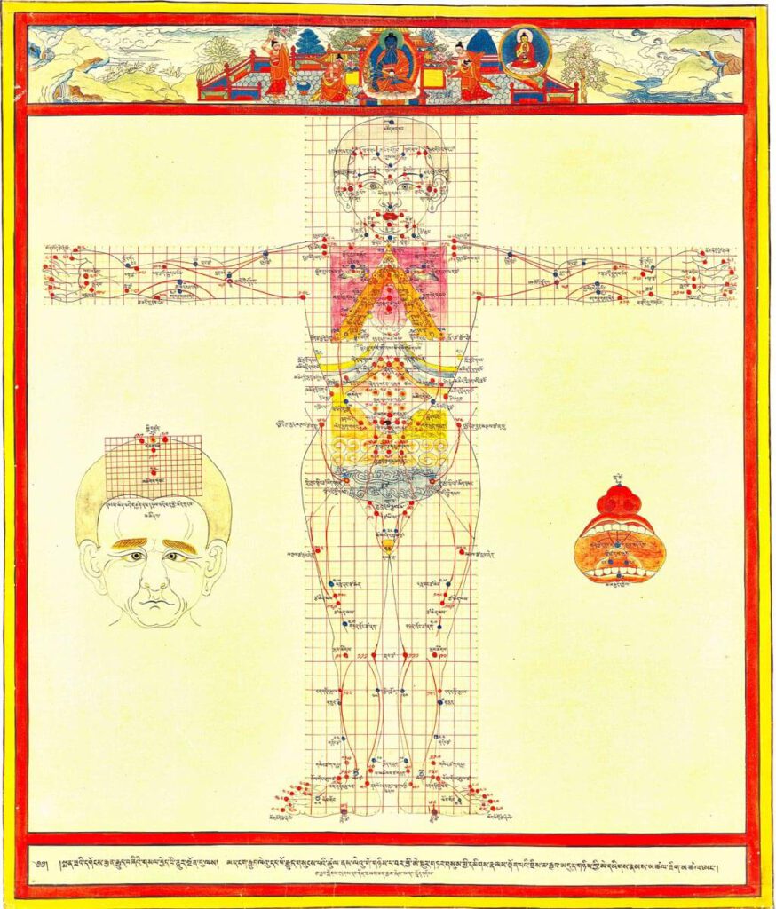
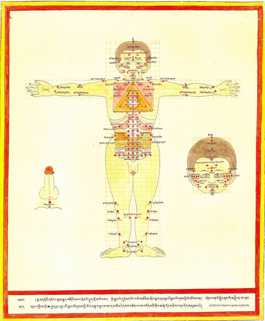
Treatment points for moxibustion, venesection, and surgery 20th century reproduction of 17th century CE painting. Held in Ulan-Ude, Republic of Buryatia, Russia. – This painting depicts the anterior view for the three types of external therapy treatment points: moxibustion, venesection, and surgical puncture. Moxibustion points are vermillion numbers and lines, venesection points are blue lines […]

Guts depicted as vulnerable part of the body
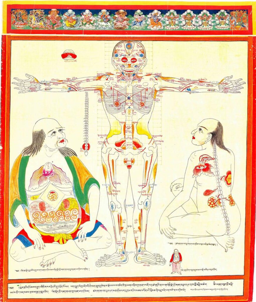
Guts depicted as vulnerable part of the body 20th century reproduction of 17th century CE painting. Held in Ulan-Ude, Republic of Buryatia, Russia. – Tibetan medicine recognizes specific vulnerable body parts to which trauma is described to be terminal or at least treated with difficulty. All critical internal organs are defined as vulnerable, the left […]

Minor connecting blood vasculature of the body including gut
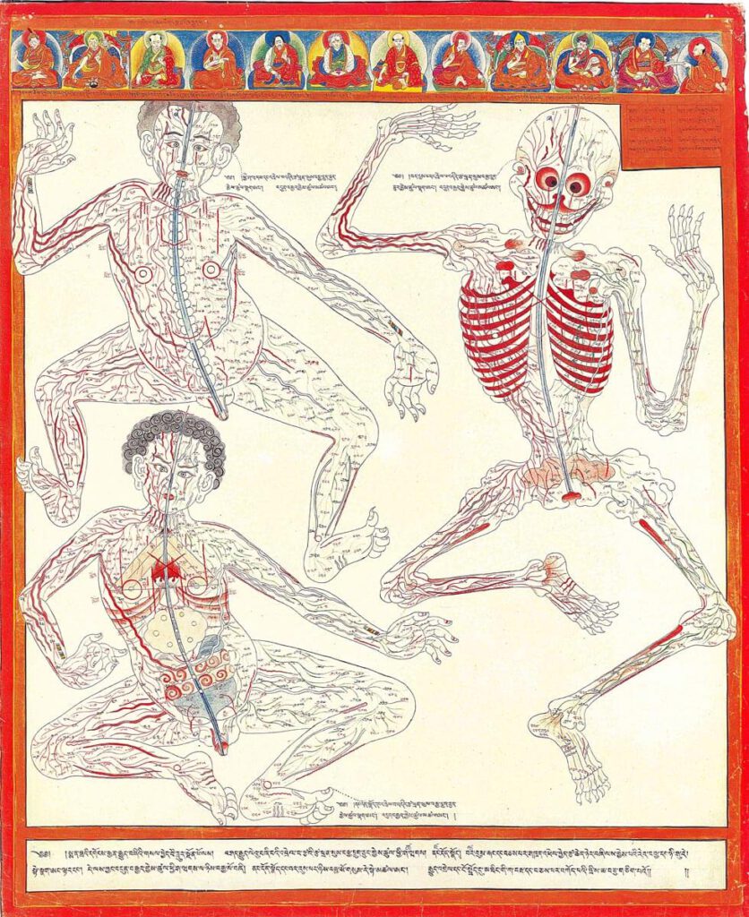
Minor connecting blood vasculature of body including gut 20th century reproduction of 17th century CE painting. Held in Ulan-Ude, Republic of Buryatia, Russia – The Four Tantras details specific enumerations of minor channels illustrated here in anatomical layout with important physiological functions. There are 360 interconnecting channels as they relate to skin and muscle tissue, […]

Major connecting blood vasculature of the body including gut with related vulnerable points
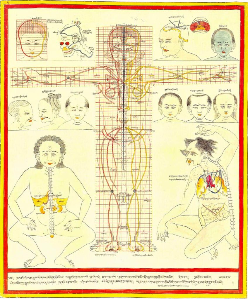
Major connecting blood vasculature of body including gut with related vulnerable points 20th century reproduction of 17th century CE painting. Held in Ulan-Ude, Republic of Buryatia, Russia. – Explicated in high detail in the traumatology section of the Four Tantras as developed from early military medical manuals, this painting depicts major blood vessels of the […]

Body metaphors including guts
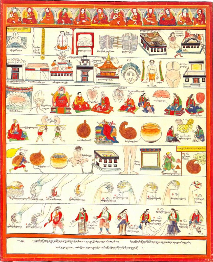
Body metaphors including guts 20th century reproduction of 17th century CE painting. Held in Ulan-Ude, Republic of Buryatia, Russia – The musculoskeletal system is likened to the architecture of a Tibetan temple, while internal organs, differentiated as solid (vital) and hollow (vessel) types, use different illustrative metaphors. The group of solid viscera include the heart, […]

Location of internal organs on modular grid
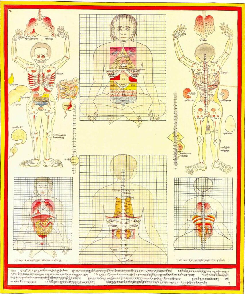
Location of internal organs on modular grid 20th century reproduction of 17th century CE painting. Held in Ulan-Ude, Republic of Buryatia, Russia. – The central figures present the anterior and posterior views of the proper positions and proportions of anatomical structures from a theoretical perspective in relation to the spine by means of the Tibetan […]

Anatomy of the gut, anterior view
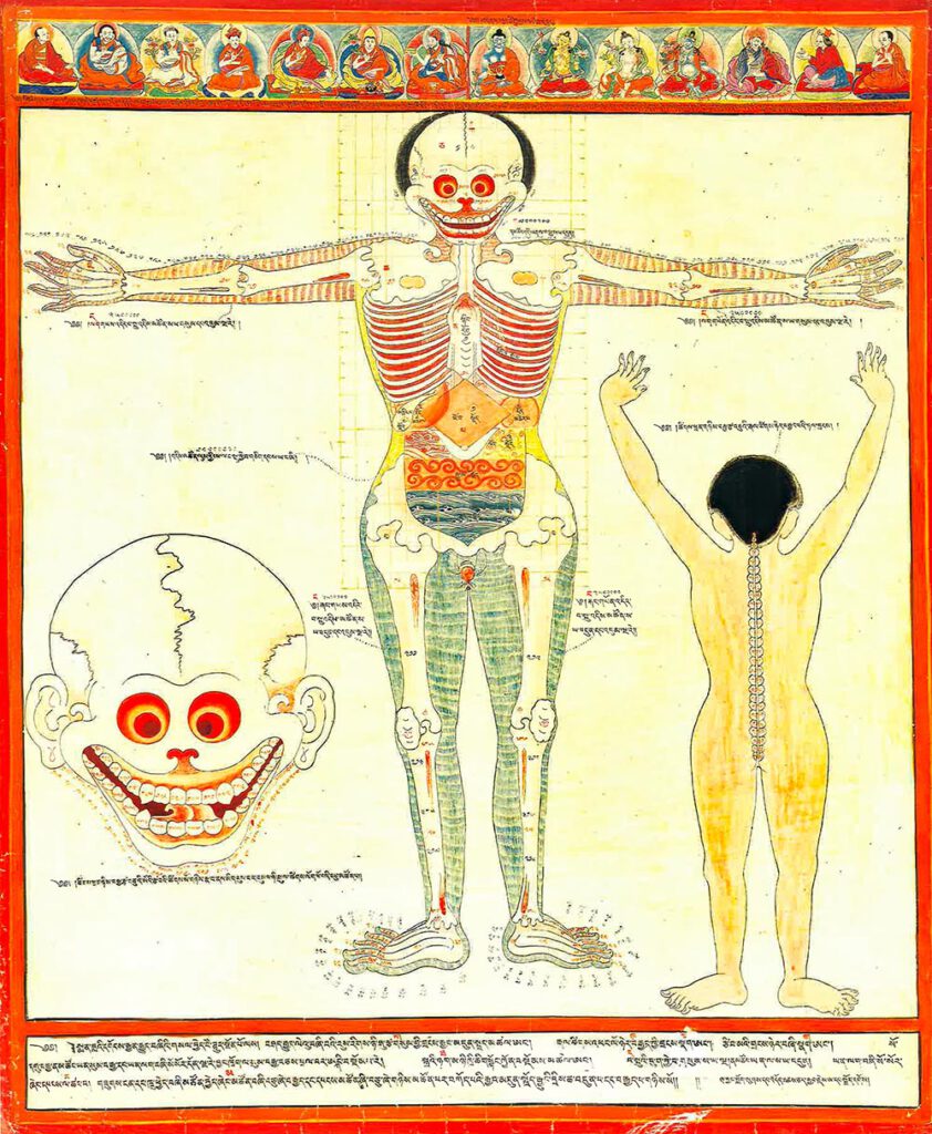
Anatomie des Darms, Vordere Ansicht — Das hochformatige Bild ist eine farbige Reproduktion aus dem 20. Farbige Reproduktion eines Gemäldes aus dem 17. Jahrhundert. In der Mitte eine aufrecht stehende Figur in der Vorderansicht abgebildet. Im linken Bildviertel ist ein Schädel dargestellt, und im rechten Bildviertel eine Rückenansicht der Figur. Teilweise sind Körperteile und innere Organe mit schwarzen Schriftzeichen versehen. Die Gestalt hat die Arme zu den Seiten ausgebreitet. Die Handflächen weisen nach vorne. Ihre Füße stehen nah beieinander, wobei die Zehen nach außen gerichtet sind. Einzelne Bestandteile des Skeletts sind detailliert dargestellt, wie zum Beispiel Fingerknochen und Fingergelenke oder der Aufbau des Kniegelenks. Flächen zwischen den Knochen und der Haut, die Fleisch und Muskeln darstellen, sind unterschiedlich farbig schraffiert. An den Armen hellbraun, um den Körper gelb und an den Beinen grün-blau. Zahnfleisch, Augenhöhlen und die Räume zwischen den Rippen leuchten in einem kräftigen Rot. In den Augenhöhlen markieren gelbe Kreise die Augen. Im leicht geöffneten breiten Mund sind die kompletten Zahnreihen sichtbar. Durch den Hals verläuft eine Röhre abwärts, die in ein auf der Spitze stehendes bräunliches Quadrat, den Magen, mündet. Um den Magen gruppieren sich weitere Organe. Auf der linken Seite des Magens befindet eine cremefarbene, tropfenförmige Form, die die Gallenblase darstellt. Rechts vom Magen befindet sich eine längliche, bauchige Form in einem ähnlichen Braunton wie der Magen, die die Milz darstellt. Unterhalb des Magens ist ein quer verlaufendes Band in Rottönen abgebildet. Es steht für den Dünndarm. Im Unterleib verläuft die Harnblase in horizontalen Wellen der Dickdarm. Die Wellen sind in Blau dargestellt. Zwei einander zugewandte Trapeze zu beiden Seiten des aufrechten bräunlichen Quadrats des Magens stellen den aufsteigenden (in leuchtend roten Wirbeln) und den absteigenden (in leuchtend gelben Wirbeln) Teil des Dickdarms dar. Die aufsteigende rote Seite ist als "Blutdarm" bekannt und steht für die Qualität des Blutes, während die gelbe Seite als "Sinbu-Darm" für die Mikroorganismen (auf Tibetisch "Sinbu" genannt) bekannt ist, die sie nährt, um Zugang zur Nahrung zu erhalten. Die obere Bildhälfte ist mit einem schwach gezeichneten Raster aus kleinen Quadraten hinterlegt. Der Schädel im unteren linken Bildviertel stellt eine Vergrößerung dar. Er entspricht im Wesentlichen dem Schädel der Gestalt. Im Unterschied zu ihm sind ausgeformte Ohrmuscheln gezeichnet. Die einzelnen Zähne sind beschriftet. Weitere Schriftzeichen verlaufen von Ohrmuschel zu Ohrmuschel entlang des Unterkiefers. Die Gestalt in Rückenansicht im rechten Bildviertel steht aufrecht mit erhobenen Armen. Ihren Hinterkopf bedecken kurze schwarze Haare. Die Körperfläche ist in Gelb gestaltet. Die Wirbelsäule mit den einzelnen Wirbeln tritt in Weiß hervor. Das Bild ist von einem strahlenden roten Band vollständig umrahmt. Eine Reihe von 14 Abbildungen von sitzenden Buddhas, Bodhisattvas und Meistern der tibetischen medizinischen Überlieferungslinie verläuft am oberen Rand des Bildes, wie bei tibetischen Gebetsfahnen. Entlang des unteren Bildrandes ziehen sich drei Schriftzeilen. Diese medizinische Darstellung kann als Teil einer ikonografischen Maltradition verstanden werden, die in buddhistischen Kulturen ihren Ursprung hat. —— Anatomy of the Gut, Anterior View — The portrait format image is a 20th-century coloured reproduction of a 17th-century painting. In the centre, an upright figure is depicted in an anterior view. The left quarter of the picture shows a skull, and the right quarter shows a posterior view of the figure. Parts of the body and internal organs are marked with black letters. The figure’s arms spread out to the sides. The palms of the hands point forward. The figure’s feet are close together with the toes pointing outwards. Individual components of the skeleton are depicted in detail, such as the finger’s bones and joints or the structure of the knee joint. Areas between the bones and the skin, representing flesh and muscles, are hatched in different colours. Light brown on the arms, yellow around the body and green-blue on the legs. Certain areas stand out in a strong red, such as the gums, eye sockets, and the spaces between the ribs. Yellow circles mark the eyes embedded within the sockets. In the slightly open and wide mouth, complete rows of teeth are visible. A tube runs down through the neck, ending in a brownish square standing on top, the stomach. Other organs are grouped around the stomach. To the left of the stomach is a cream-coloured tear-dropped shape gall bladder. To the right of the stomach is a bulbous oblong shape, in a similar shade of brown to the stomach, the spleen. Below the stomach, a transverse band is depicted in shades of red. It represents the small intestine. In the lower abdomen, the urinary bladder runs in horizontal waves. The waves are depicted in blue. Two bi-facing trapezoids on either side of the upright brownish square of the stomach represent the ascending (in bright red swirls) and descending (in bright yellow swirls) parts of the large intestine. The ascending red side is known as the 'blood colon' related to nourishing blood quality and the yellow side is known as the 'sinbu colon' for the microorganisms (known as 'sinbu' in Tibetan) that it nourishes to access nutrition. The upper half of the picture has a faintly drawn grid of small squares. The skull in the lower left quarter of the picture represents an enlargement. Essentially, it corresponds to the skull of the figure. In contrast to it, shaped auricles are drawn. The individual teeth are labelled. Further characters run from auricle to auricle along the lower jaw. The figure in the posterior view in the right quarter of the image stands upright with raised arms. Short black hair covers the back of the head. The body is painted in yellow. The spine with individual vertebrae stands out in white. The picture is completely framed by a radiant red band. A row of 14 images of seated Buddha, bodhisattva, and Tibetan medical lineage masters runs along the upper edge of the picture, as in Tibetan prayer flags. Three lines of script run along the lower edge of the picture. This medical representation can be understood as part of an iconographic painting tradition that originated in Buddhist cultures.

Anatomy of the gut, anterior view 20th century reproduction of 17th century CE painting. Held in Ulan-Ude, Republic of Buryatia, Russia. – This painting displays an enumeration of body part components (e.g., bones, vertebrae, joints, etc.) and diagrams the proper associated relational measures in anterior view using a quadrant grid. The abdominal quadrant covers 12×8 […]