Attempts to access the interior of the body via orifices had already been made since antiquity, with particular attention being paid to the oral, genital, urinary and excretory organs. Phillip Bozzini (1773-1809) was also active in this field, seeking, as he put it, to “illuminate the inner cavities of the living animal body”. In 1806, he was able to present his “Lichtleiter”, in which the light from a candle was reflected into the interior of the urinary bladder via a mirror [1].
Thus the basic idea for the cystoscope (literally “bladder viewer”) was developed.
The urologist Maximilian Nitze (1848-1906) took up this idea at the end of the 19th century and presented his cystoscope in 1879, which allowed direct illumination and examination of human and animal body cavities by means of electric light [2].
However, there was now the problem of visual fixation. The examination was painful and patients could hardly be expected to wait until an artist had drawn what they had seen. At that time, a photograph had the disadvantage that, first, it only allowed black-and-white images and, second, it also could not be taken in a matter of seconds. But was the black-and-white image really a disadvantage? Wasn’t the black-and-white image clearer in its simplicity, and thus more evident? Wasn’t the subjective coloring of the illustrators distorting?
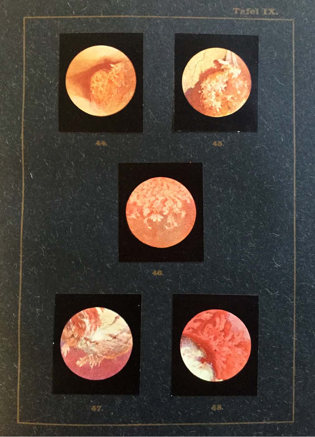
This sparked off a debate in which the urologist Otto Kneise defended the colored hand-painted pictures. He praised an Atlas of Cystoscopy published by him in 1908 as a collection of “good pictures, corresponding to the natural conditions also in their light and color effects”. Then again, he spoke of the difficulty of “reproducing cystoscopic images in color” [3].
In particular, he pointed out that colors appear quite different in artificial light than in sunlight, that the different light intensities or shadowing in cystoscopy have considerable influence on color perception.
The central problem was creating images “true to nature” and evidential value. It was disputed whether colorfulness or photographic objectivity was better suited to achieve evidential effects. The advantages of colorfulness were relativized by doubts about rendering it “true to nature” through the subjectivity of the artistic process. In contrast to this stood the “objectivity” of photography, which in turn was afflicted with the shortcoming of colorlessness. In fact, the latter, the shortcoming of colorlessness, obviously weighed as a more serious disadvantage, because almost all contemporary textbooks and atlases on cystoscopy opted for the colored variant [4].
1 Reuter, Matthias (2006): Phillip Bozzini (1773-1809). Der endoskopische Idealist. Der Urologe 45, p. 1084-1091.
2 Herr, Harry W. (2006): Max Nitze, the Cystoscope and Urology. The Journal of Urology 176, p. 1313-1316.
3 Kneise, Otto (1908): Handatlas der Cystskopie. Gebauer-Schwetschke: Halle a. Saale.
4 Martin, Michael and Fangerau, Heiner: Evidenzen der Bilder. Visualisierungsstrategien in der medizinischen Diagnostik um 1900. Steiner, Stuttgart 2021.