The images presented here were produced around 1900. The context is that of Western Medicine, which was transformed during the 19th century into a science oriented towards modern chemistry, physics, and mathematics rather than towards history and philosophy.
In the hierarchy of the senses, seeing has occupied a special position in Western modernity at least since the Enlightenment [see 1, 2]. Seeing something suggested truth and knowledge. What was in the dark, by contrast, was invisible, indistinct, and the subject of speculation. In the context of the establishment of a scientifically and technically oriented medicine, efforts were made to eliminate subjective elements and to elevate the object of inquiry to the status of an object that could be scientifically observed and documented. For example, images that made the previously invisible visible, or that clarified the visible, were now used to elevate facts to the status of an object of scientific justification. The aim was thus to create images that were as objective as possible, ideally using technical means to eliminate the fallibility and inadequacy of the human senses.
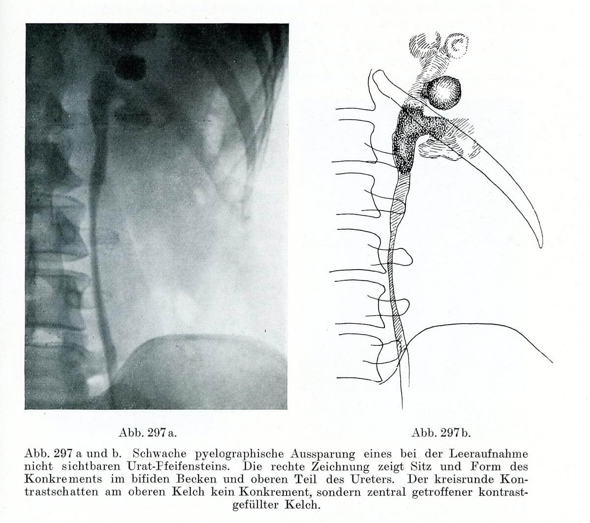
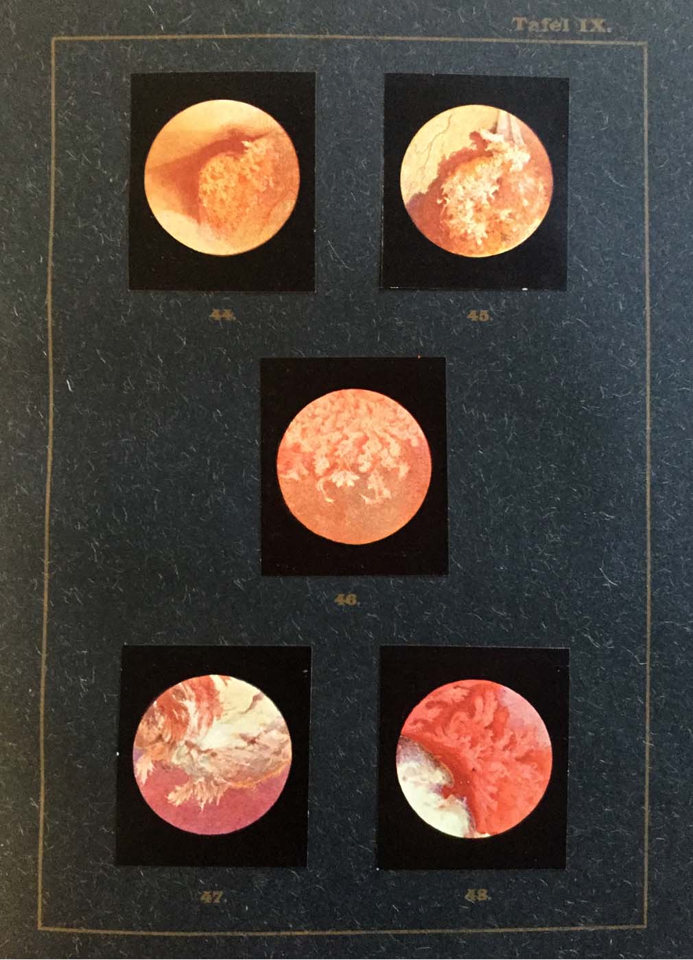
In medical diagnostics, pictorial documentation caused problems. The conservation of images appeared to be necessary for monitoring the course of a disease, for teaching purposes, and for the development of diagnostic theory. When it came to signs that could be perceived visually, however, linguistic description did no justice to what was visible, and drawings offered only limited diagnostic certainty, being at most a substitute for direct inspection. Developing technologies of producing images independent of subjective senses or skills accordingly became an objective of medical imaging. Examples include endoscopy and the use of X-ray technology as diagnostic procedures.
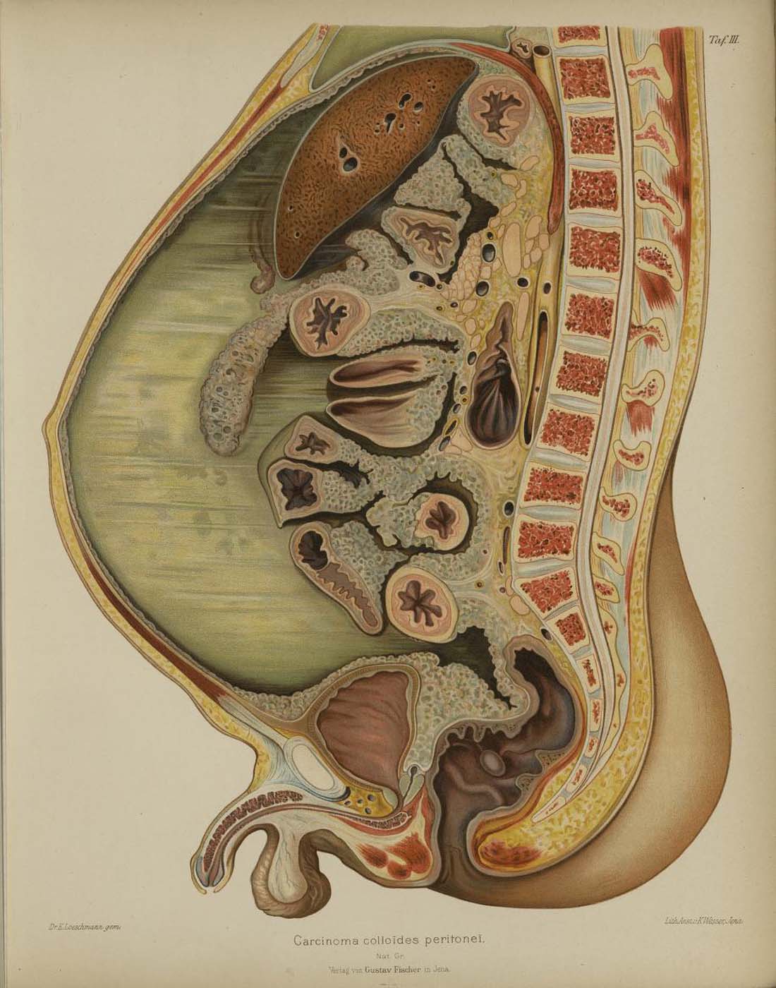
The production of images from the interior of a living patient’s body was particularly difficult, and at the same time spectacular. Here elaborate techniques had to be developed to make hidden organs and structures, such as the guts, both visible and diagnostically meaningful. What could be seen had to be made accessible to scientific discourse. To this end, countless manuals and textbooks, intended to serve as reference works for the “normal pathological”, were published in the late 19th and 20th centuries. But the teaching of conceptions of normality, notions of pathology, and learning to read visualizations brought with them a central problem, by contradicting the demand for an objective, unaltered immediacy of diagnostic signs. A susceptibility to deception and subjective influences was a problem for technically produced images from the interior of the human body. For diagnostic imaging, this resulted in a tense ambivalence: interior body parts were directly accessible to the sense of sight, but at the same time, what was seen was bound to indirect procedures of technical reproduction. The simultaneous immediacy and mediated nature of images from within the body became the subject of ongoing contemporary discussion, and resulted in attempts to develop technologies producing self-evident images [1].
1. Martin M., and Fangerau H. (2011): “Töne sehen? Zur Visualisierung akustischer Phänomene in der Herzdiagnostik”, NTM Zeitschrift für Geschichte der Wissenschaften, Technik und Medizin 19(3): 299–327.
2. Fangerau H. (2017): Bilder, Zahlen und die Medizin der Zukunft: Das “Als ob” als Philosophie der Prävention. In: Kessler, S., Fangerau, H., and Wiesing, U. (eds.): Präventionsentscheidungen: Zur Geschichte und Ethik der Gesundheitsvorsorge im 21. Jahrhundert. Frommann-Holzboog, Stuttgart, pp. 59–94.