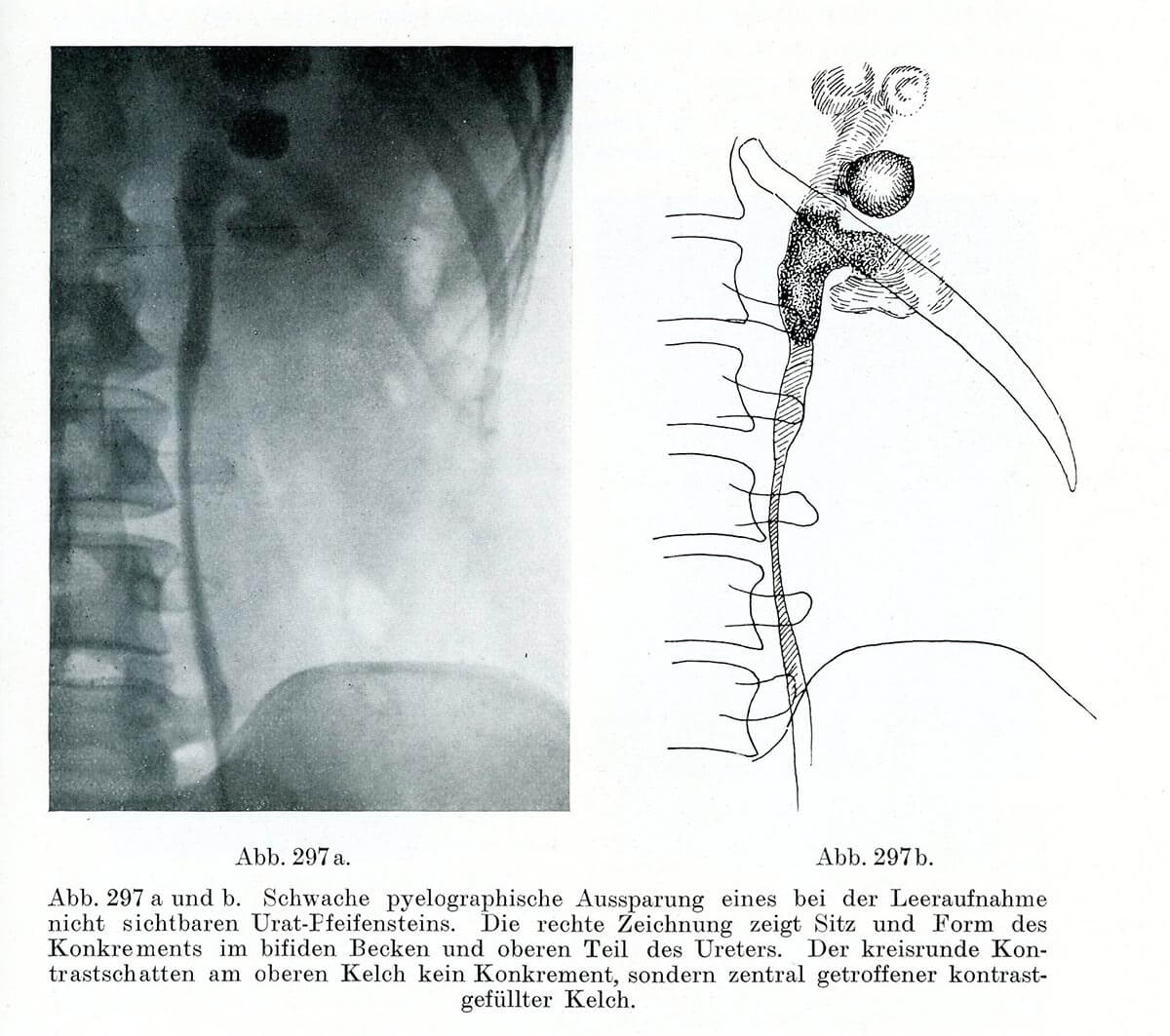
Leopold Casper und Edwin Picard (1930) Lehrbuch der urologischen Diagnostik. Thieme, Leipzig, S. 397
Right after the discovery of X-rays, in the late 19th century, innumerable attempts were made to make this novel technique useful for medicine. While the radiological detection of bones quickly became a routine procedure, it was necessary to first develop specific procedures for the radiographic visualization of soft tissue organs and cavities. The problem was that only structures that absorbed X-rays were visible on the negative film. Rays simply passed through soft tissues. The film remained black. The testing of various means used to generate contrast (e.g. iodine) proved to be risky, which is why their use was initially controversial [1,2].
1. Dommann M (1999) „Das Röntgen-Sehen muss im Schweisse der Beobachtung gelernt werden.” Zur Semiotik von Schattenbildern. Traverse, Zeitschrift für Geschichte. Wissenschaft, die Bilder schafft 6:114-130
2. Martin M and Fangerau H (2012) “Durchsichtbarkeitsregime: Zur Semiotik radiographischer Bilder in der urologischen Diagnostik”, Der Urologe 51: 1450–1458