Representation of a woman with the distribution of the blood vessels and the internal organs (f. 15v)
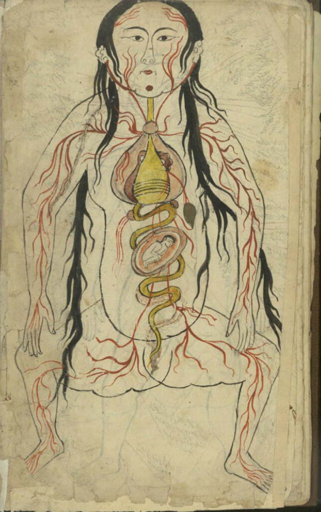
Representation of a woman with the distribution of the blood vessels and the internal organs (f. 15v) Manṣūr ibn Ilyās (fl. 14th c.), Tashrīḥ-i badan-i insān. Images from: Teheran MS Majlis 7430. Undated. This image represents the heart, lungs, liver, esophagus/larynx, stomach, intestines, kidneys, and spleen). As in the male representation, the organs involved in […]
Representation of a man with the distribution of the blood vessels and the internal organs (f. 13v)
Representation of a man with the distribution of the blood vessels and the internal organs (f. 13v) Manṣūr ibn Ilyās (fl. 14th c.), Tashrīḥ-i badan-i insān. Images from: Teheran MS Majlis 7430. Undated. The illustration depicts the heart, lungs, liver, esophagus/larynx, stomach, intestines, kidneys, and spleen. The organs involved in digestion are coloured in yellow. […]
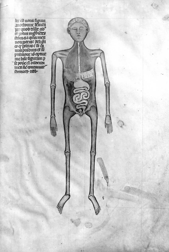
Illustration accompanying De Arte Physicali et de Cirurgia
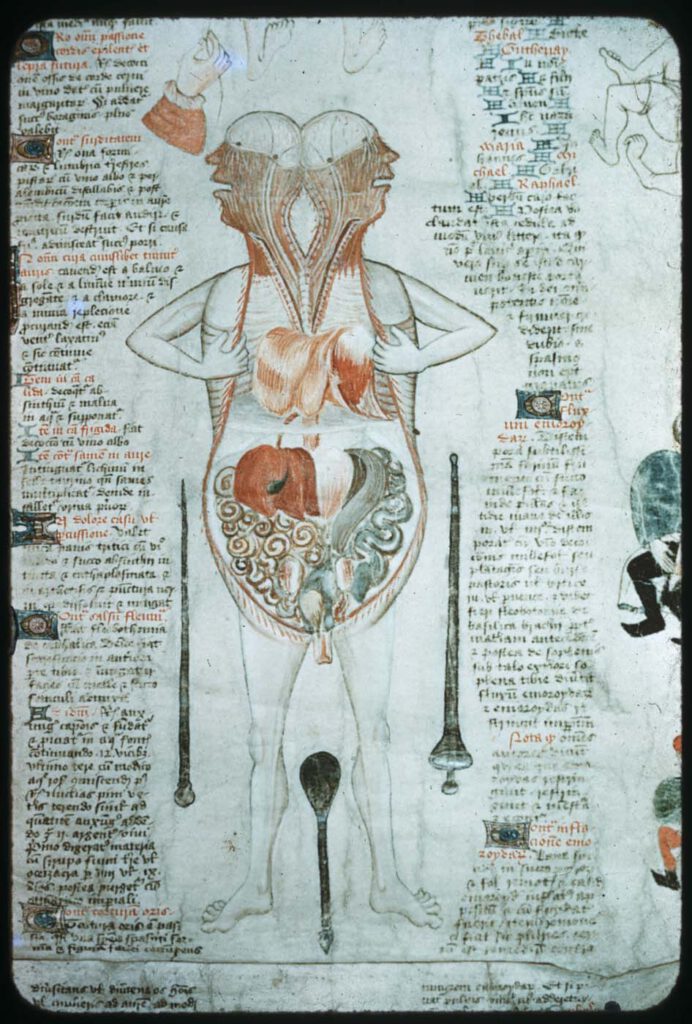
Illustration accompanying De Arte Physicali et de Cirurgia, attributed to John Arderne Stockholm, Kungliga Biblioteket X.118, fol. 6v/6r; c. 1430 – While the frontal approach to anatomical illustration exhibited in the Five/Nine-Figure Series and Guido da Vigevano’s work is most dominant, an alternative, sagittal approach emerged in the medieval period and survives in a few […]
Guido da Vigevano’s Anathomia, Figure 8
Guido da Vigevano’s Anathomia, Figure 9 (1345) Guido da Vigevano, Anathomia, 1345CC BY-NC-SA 4.0 – Although human dissection began to be systematically practiced in Italy in the early 14th century, it was not immediately embraced across Europe. This prompted Guido da Vigevano to produce a series of seventeen illustrations included in his Book of Notable […]
Anatomical figures
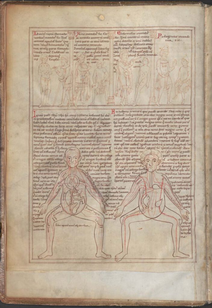
Anatomical figures (Munich, Bayerische Staatsbibliothek, Clm 13002, fol. 2v; 1165 CE) Photo: Bayerische Staatsbibliothek MünchenCC BY-NC-SA 4.0 – These figures offer the earliest example of the so-called Five-Figure Series (Fünfbilderserie), which appears in medical manuscripts from across the medieval world, with Latin, Provençal, Arabic, and Persian versions surviving. The series, which likely originated in Late […]
Illustrations of the uterus
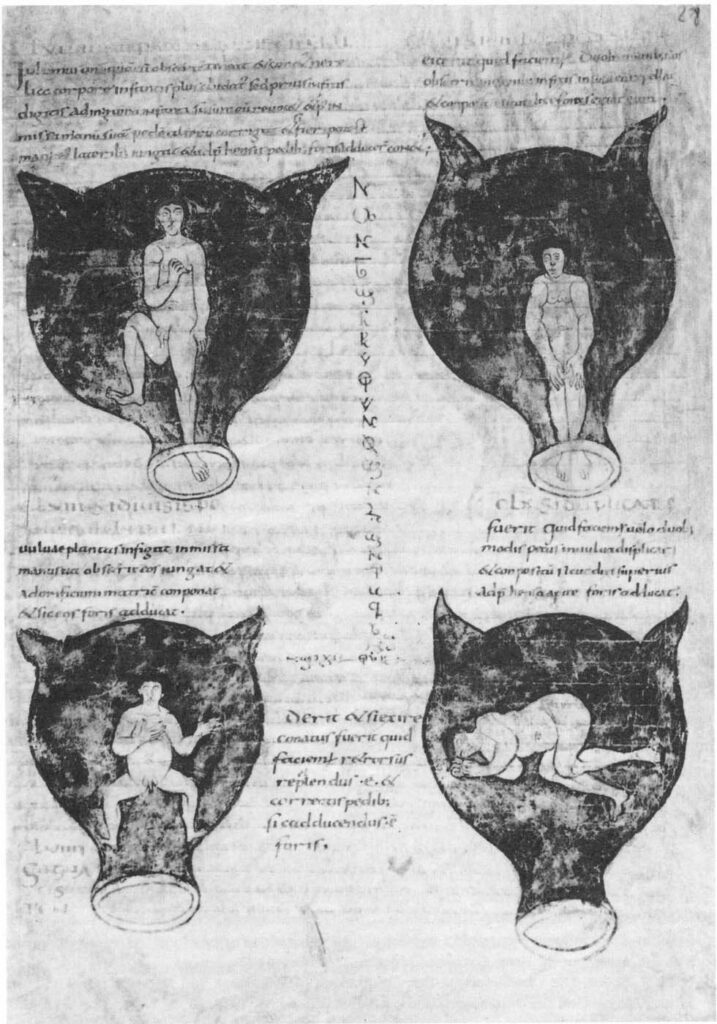
Illustrations of the uterus from Muscio’s epitome of Soranus’ Gynecology Brussels, Bibliothèque Royale MS 3714, fol. 28r; c. 900 CECC0 1.0 Universal (CC0 1.0) – These illustrations of various positions of the fetus in the womb accompany Muscio’s Late Antique Latin summary of Soranus’ 2nd century Greek work on gynecology. The 9th/10th century manuscript pictured […]
Apollo the Python-Slayer or Cleveland Apollo
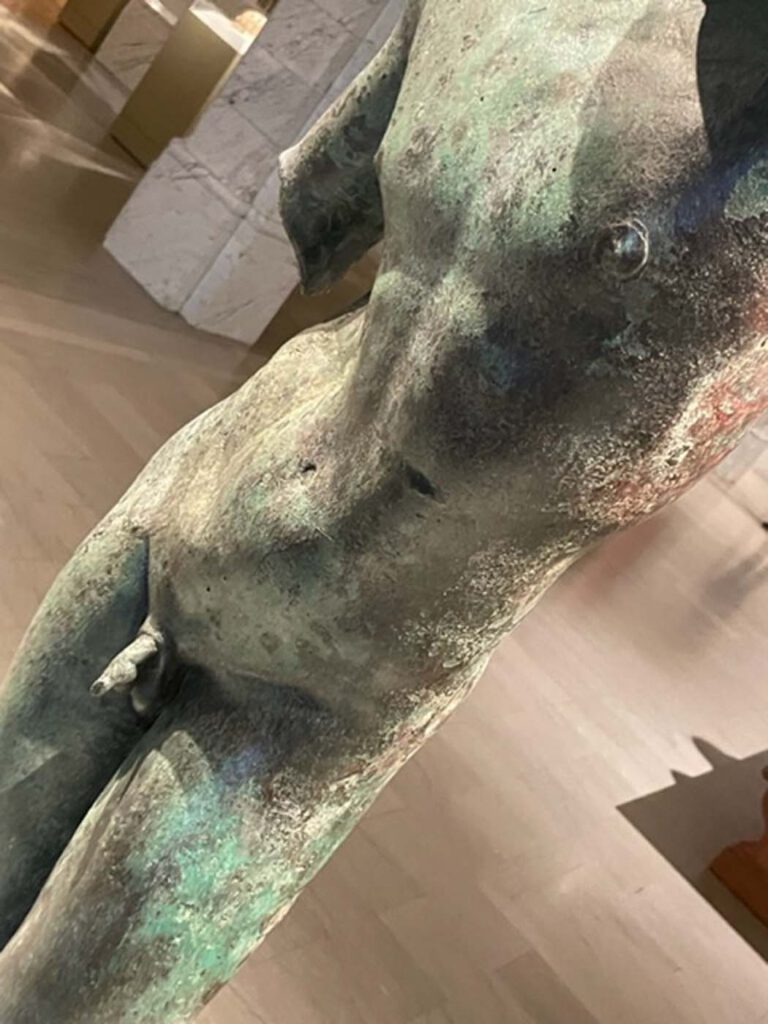
Apollo the Python-Slayer or Cleveland Apollo Hellenistic (?) Courtesy of the Cleveland Museum of Art. Museum Number 1839,0214.51 – The celebrated Cleveland Apollo is one of the most famous works attributed to the fourth-century Attic sculptor Praxiteles. Many later marble copies survive, but this bronze classical piece came to public attention only in 2004 when […]
Grotesque figurine
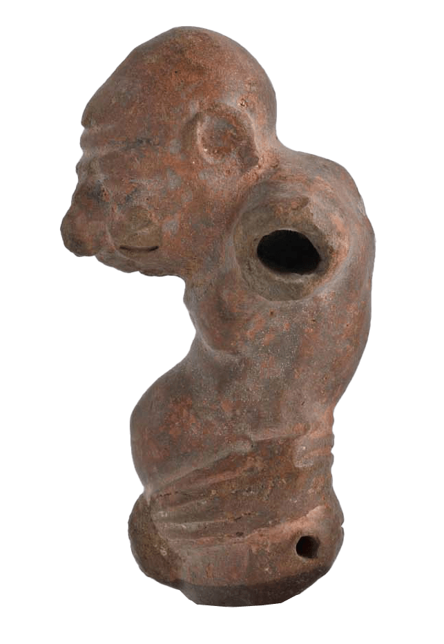
Grotesque terracotta figurine 3rd century BCE–3rd century CE (?) © 2023, Benaki Museum, Athens. Benaki Museum inv. No 12793 – This Hellenistic terracotta, part of the Benaki Museum collection in Athens, depicts with hyper-realistic traits an old, and/or ‘disabled’ man with a protruding pot belly, achieving a grotesque effect and signalling a pathology while offering […]
Marble tombstone of an Athenian physician
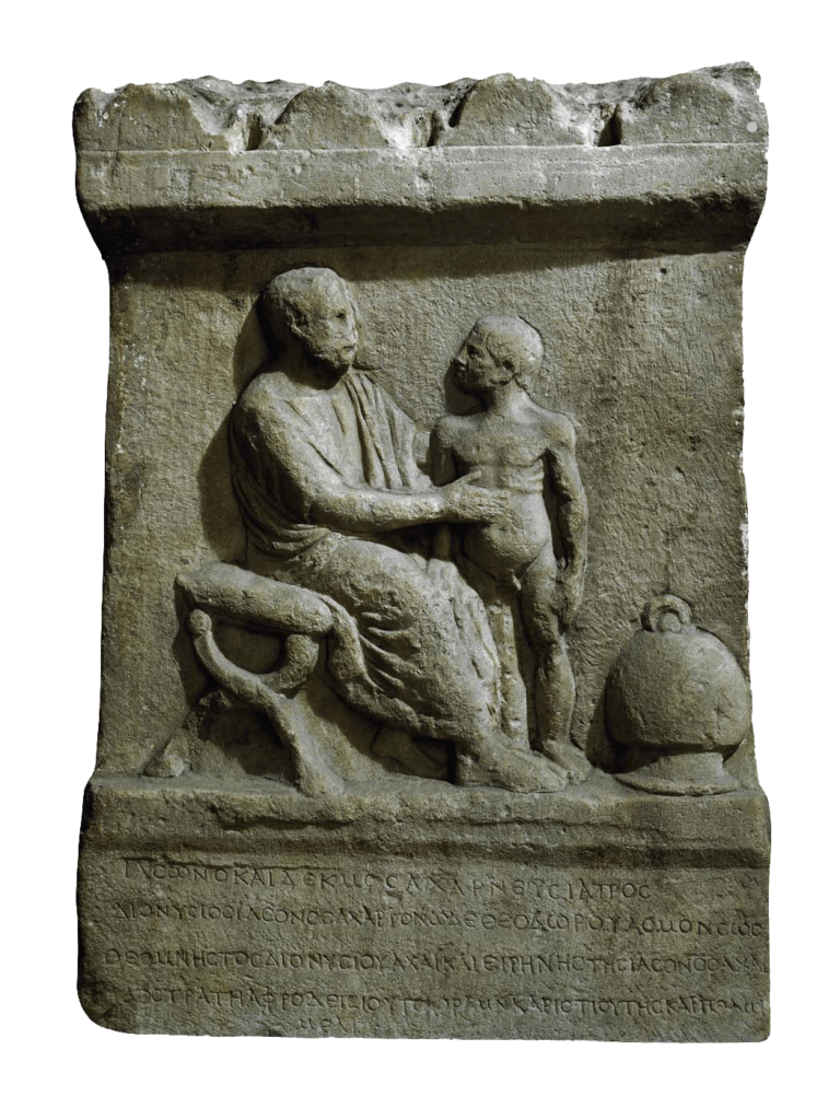
Athens, 2nd century CE