Blasengeschwülste, Fig. 44-46: Ansichten eines Papilloms, Fig. 47-48: Ansichten eines Tumors (Kneise 1908: Tafel IX)
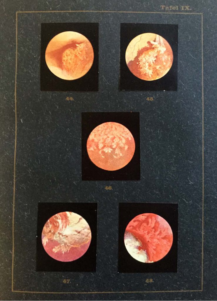
Blasengeschwülste, Fig. 44-46: Ansichten eines Papilloms, Fig. 47-48: Ansichten eines Tumors (Kneise 1908: Tafel IX) Kneise, Otto (1908): Handatlas der Cystskopie. Gebauer-Schwetschke: Halle a. Saale. — Attempts to access the interior of the body via orifices had already been made since antiquity, with particular attention being paid to the oral, genital, urinary and excretory organs. […]
Lehrbuch der urologischen Diagnostik
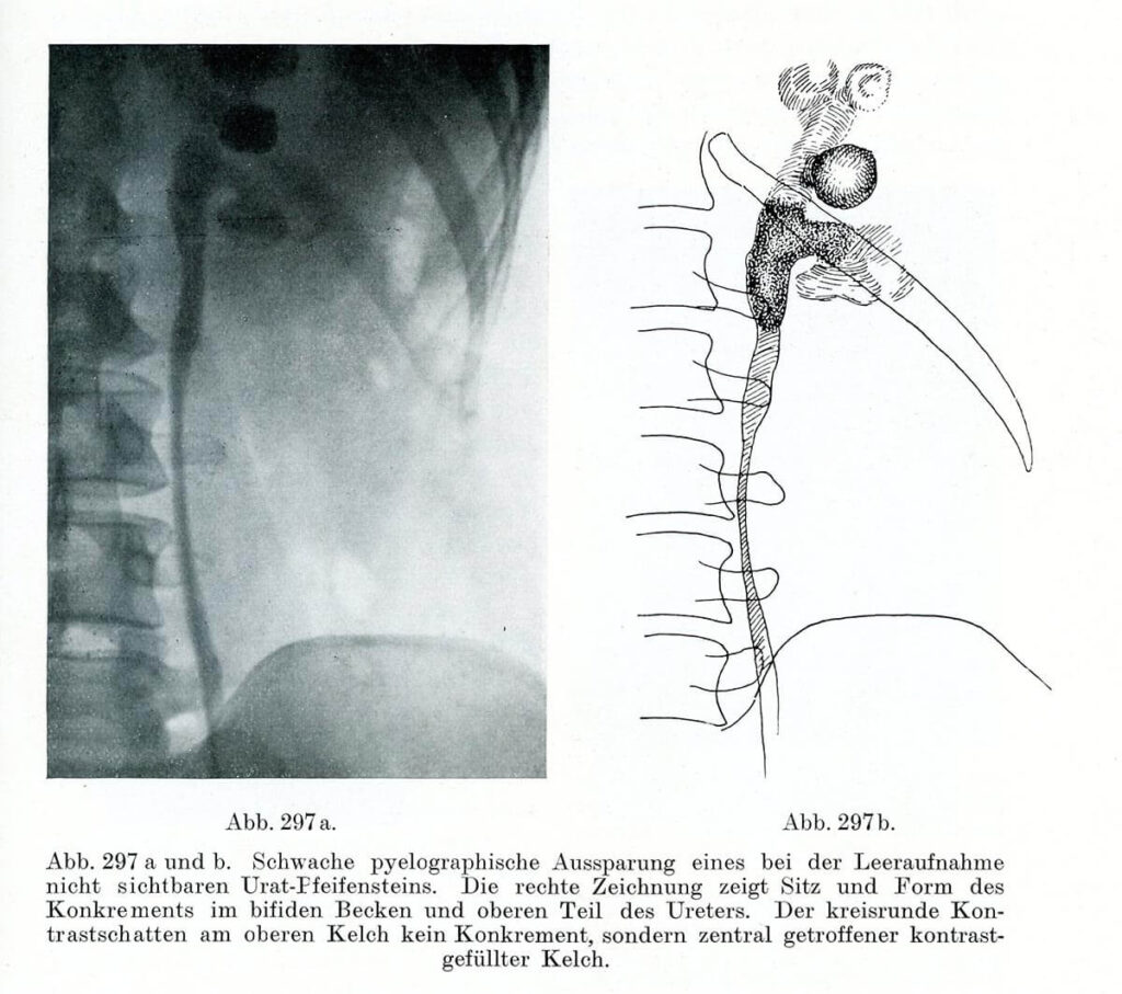
Pyeolographische Darstellung eines Konkrements im Ureter Leopold Casper und Edwin Picard (1930) Lehrbuch der urologischen Diagnostik. Thieme, Leipzig, S. 397 – Right after the discovery of X-rays, in the late 19th century, innumerable attempts were made to make this novel technique useful for medicine. While the radiological detection of bones quickly became a routine procedure, […]
Carcinoma colloides peritonei

Carcinoma colloides peritonei From: Emil Ponfick: Topographischer Atlas der Medizinisch-Chirurgischen Diagnostik / Topographic atlas of medico-surgical diagnosis, G. Fischer. Jena 1900-1905 Artist: Dr. Emil Löschmann, Breslau — The attempts of the German pathologist Emil Ponfick, who in 1901 attempted to reduce the gap between drawing and original object, show how objectives and implementation should be brought together […]