Anonymous, K. Sharḥ al-Maqāma al-Ṣalāḥiyya fī l-Khayl wa-al-Bayṭāra
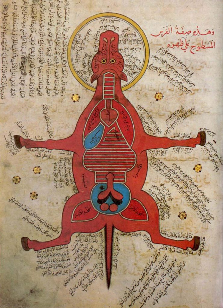
Anonymous, K. Sharḥ al-Maqāma al-Ṣalāḥiyya fī l-Khayl wa-al-Bayṭāra (Commentary on the Maqāma Ṣalāḥiyya on Horses and Venterinary) Istanbul University Library MS 4689 – Egypt, 15th c. – Hyppiatry has a long tradition in Islamic literature. Works dealing with horses may adopt different literary genres and cover a wide range of disciplines, from veterinary sciences to […]
Representation of a woman with the distribution of the blood vessels and the internal organs (f. 15v)
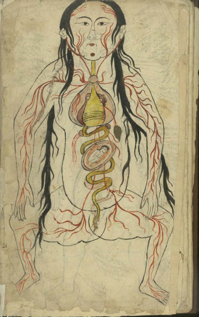
Representation of a woman with the distribution of the blood vessels and the internal organs (f. 15v) Manṣūr ibn Ilyās (fl. 14th c.), Tashrīḥ-i badan-i insān. Images from: Teheran MS Majlis 7430. Undated. This image represents the heart, lungs, liver, esophagus/larynx, stomach, intestines, kidneys, and spleen). As in the male representation, the organs involved in […]
Representation of a man with the distribution of the blood vessels and the internal organs (f. 13v)
Representation of a man with the distribution of the blood vessels and the internal organs (f. 13v) Manṣūr ibn Ilyās (fl. 14th c.), Tashrīḥ-i badan-i insān. Images from: Teheran MS Majlis 7430. Undated. The illustration depicts the heart, lungs, liver, esophagus/larynx, stomach, intestines, kidneys, and spleen. The organs involved in digestion are coloured in yellow. […]
Illustration depicting a right lateral view of the body (f. 72r)
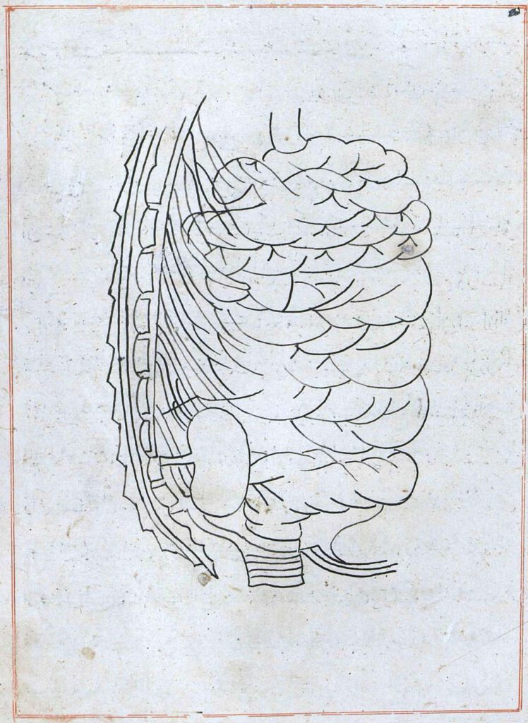
Illustration depicting a right lateral view of the body (f. 72r) Tansūqnāmah-i Īlkhān dar funūn-i ʿulūm-i Khatāʾī (Ilkhan’s Treasure Book on the Branches of the Chinese Sciences). Istanbul MS Aya Sofya 3596. Tabriz, 1313. The image closes the sixth section. It shows the spine, the right kidney, the intestines, and the bladder. – Read the […]
Frontal illustration of the small and large intestines and the bladder (f. 69v)
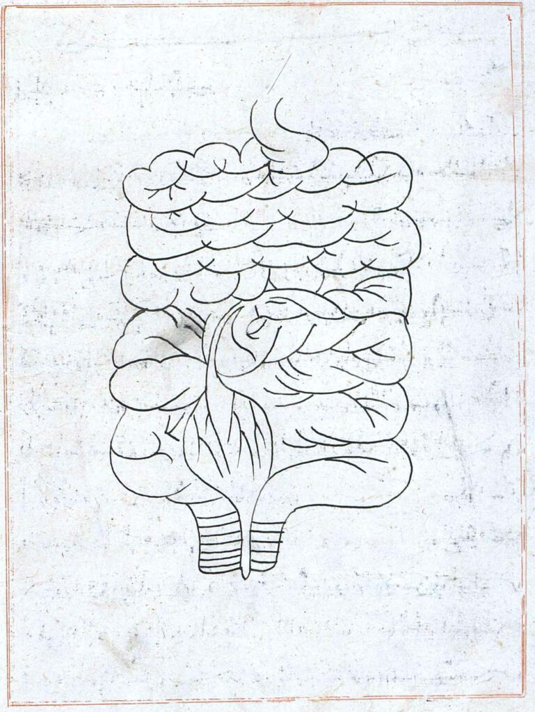
Frontal illustration of the small and large intestines and the bladder (f. 69v) Tansūqnāmah-i Īlkhān dar funūn-i ʿulūm-i Khatāʾī (Ilkhan’s Treasure Book on the Branches of the Chinese Sciences). Istanbul MS Aya Sofya 3596. Tabriz, 1313. This illustration occurs right before the beginning of the sixth section of the book (f. 70r) describing the connection […]