Comparative Guts

Search
Close this search box.

Lehrbuch der urologischen Diagnostik
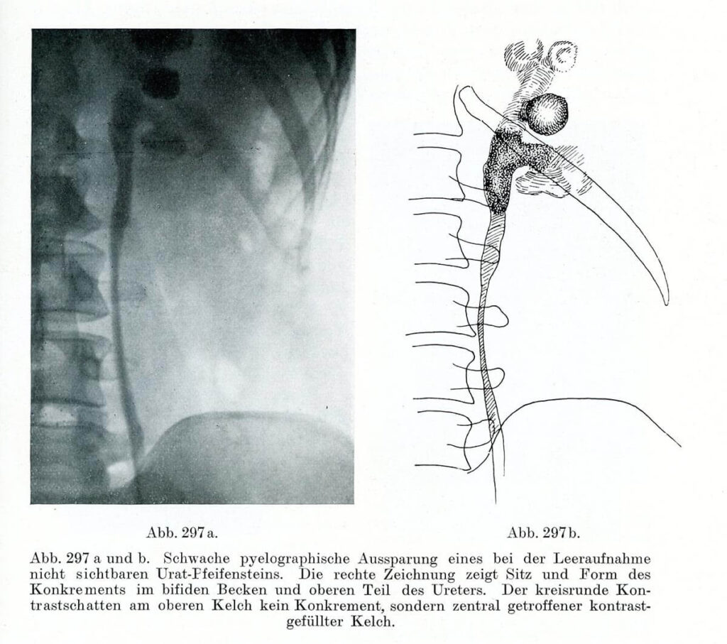
Pyeolographische Darstellung eines Konkrements im Ureter Leopold Casper und Edwin Picard (1930) Lehrbuch der urologischen Diagnostik. Thieme, Leipzig, S. 397 – Right after the discovery of X-rays, in the late 19th century, innumerable attempts were made to make this novel technique useful for medicine. While the radiological detection of bones quickly became a routine procedure, […]

Carcinoma colloides peritonei

Carcinoma colloides peritonei Die farbige Abbildung zeigt einen vertikalen Querschnitt der Bauchhöhle mit ihren Organen und der Wirbelsäule. Sie füllt die gesamte Seite aus. Auf dem schmalen Rand rechts oben steht in lateinischen Buchstaben Taf. 3. Am unteren Rand ist zentriert in blasser gedruckter Schrift der Titel der Zeichnung lesbar. Die Darstellung beginnt am oberen Seitenrand unterhalb des Brustkorbs und endet unten mit dem Po und männlichen Geschlechtsorganen. Rechts ist sie durch die Wirbelsäule begrenzt, links durch den stark vorgewölbten Bauch. Umgeben wird die Abbildung von einen beige-gelblichen Rand. Die Wirbelsäule schwingt im Lendenbereich etwas nach links. Die einzelnen Wirbel sind in einem hellem Gelb dargestellt, rote Pünktchen dicht nebeneinander markieren ihr Inneres. In Hellblau erscheinen die Räume dazwischen. Nach links folgt die Abbildung der Bauchhöhle mit ihren Organen. Sie sind mehrheitlich in einem blassen Rot gezeichnet. Blassgelbe Umrandungen konturieren sie nach außen. Alle Organe sind dazu mehr oder weniger von grauer Farbe umgeben. Sie erscheint wie ein Belag, dessen Struktur an Kaviar erinnert. Im oberen Drittel sticht eine ovale braune Fläche hervor, auf der unterschiedlich große dunkle Punkte erscheinen. Darunter, nah an der Wirbelsäule befindet sich eine längliche, vertikal verlaufende Farbfläche in Gelb, deren Enden sich verjüngen. Darin ist ein dunkelbraunes Oval abgebildet. Der Raum hinter der Wölbung der Bauchdecke links ist mit blasser grün-grauer Farbe ausgemalt. Diese Abbildung stammt aus dem Topographischen Atlas der Medizinisch-Chirurgischen Diagnostik, der vom Pathologen Emil Ponfick zwischen 1900 und 1905 zusammengestellt wurde. Unterstützt wurde er dabei von Dr. Emil Löschmann, einem Maler und Grafiker aus Breslau. — — Carcinoma colloides peritonei The coloured illustration shows a vertical cross-section of the abdominal cavity with its organs and spine. It fills the entire page. On the narrow margin in the upper right corner, “Taf. 3” is written in Latin letters. The title of the drawing can be read at the lower margin, centred in a pale printed script. The illustration begins at the top of the page below the ribcage and ends at the bottom with the buttocks and male sexual organs. On the right, it is limited by the spine, and on the left by the strongly bulging abdomen. The figure is surrounded by a beige-yellowish border. The spine swings slightly to the left in the lumbar region. The individual vertebrae are depicted in light yellow, red dots close together mark their interior. The spaces between them appear in light blue. To the left follows the illustration of the abdominal cavity with its organs. The majority of them are drawn in pale red. Pale yellow borders outline them on the outside. All organs are more or less surrounded by a grey colour. It appears like a coating whose structure is reminiscent of caviar. In the upper third, an oval brown area stands out, on which dark dots of different sizes appear. Below it, close to the spine, is an elongated vertically running coloured area in yellow, the ends of which taper off. A dark brown oval is depicted within it. The space behind the bulge of the abdominal wall on the left is painted in a paler green-grey colour. This illustration is from the "Topographical Atlas of Medico-Surgical Diagnostics," compiled by the pathologist Emil Ponfick between 1900 and 1905. He was assisted by Dr. Emil Löschmann, a painter and graphic artist from Breslau.

Carcinoma colloides peritonei From: Emil Ponfick: Topographischer Atlas der Medizinisch-Chirurgischen Diagnostik / Topographic atlas of medico-surgical diagnosis, G. Fischer. Jena 1900-1905 Artist: Dr. Emil Löschmann, Breslau — The attempts of the German pathologist Emil Ponfick, who in 1901 attempted to reduce the gap between drawing and original object, show how objectives and implementation should be brought together […]