The Gut / Der Darm
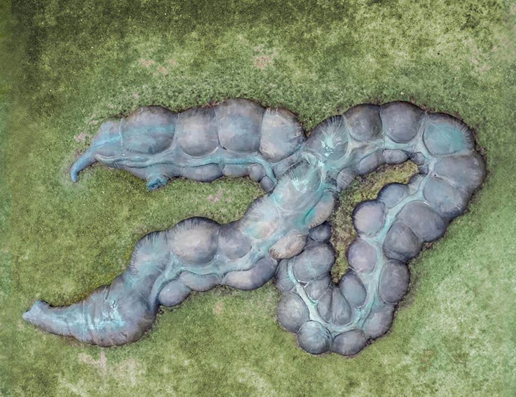
The Gut / Der Darm Anetta Mona Chisa & Lucia Tkacova public sculpture, Schillerplatz, Chemnitz (part of the project Gegenwarten | Presences), photographed by the artist – The Gut is the organ that renders Marx’s body in the city that holds the largest sculpture of Marx’s head in the world – Karl-Marx-Monument. The actual head […]
Henry Gray (anatomist) and Henry Vandyke Carter (artist), Fig. 1005, Superior and inferior duodenal fossæ.
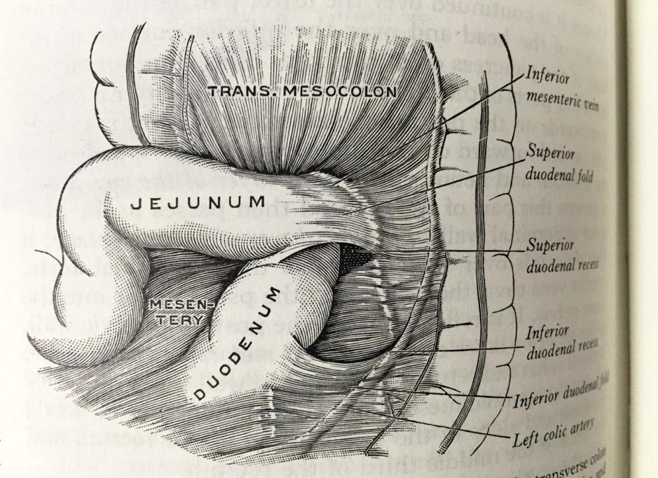
Henry Gray (anatomist) and Henry Vandyke Carter (artist), Fig. 1005, Superior and inferior duodenal fossæ. Gray’s Anatomy Descriptive and Applied. A New American Edition (1913), p. 1265.Private collection: Nina Sellars. Photographer: Nina Sellars. – Giving thoughtful attention to the physical qualities of the classical anatomical atlases brings our awareness not only to their content and […]
Wangechi Mutu, Histology of the Different Classes of Uterine Tumors (12 works) (2004–05)
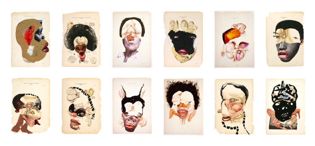
Wangechi Mutu, Histology of the Different Classes of Uterine Tumors (12 works) (2004–05) Series of twelve collages on found printed medical illustrations, including printed paper, glitter, ink, adhesive tape and fur. © Wangechi Mutu. Courtesy of the artist – Wangechi Mutu works expansively across sculpture, film, collage, performance, drawing, and painting. In dialogue with Afrofuturism, […]
Mary Reid Kelley with Patrick Kelley, This is Offal (2015)
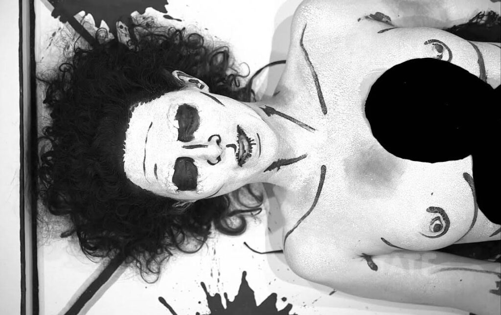
Mary Reid Kelley with Patrick Kelley, This is Offal (2015) Live performance streamed via internet, 8 min., Tate Modern, November 19, 2015; Photo credit: Patrick Kelley. Courtesy of the artists A video of the performance is available here – Mary Reid Kelley works at the boundary between performance, painting, and poetry. She crafts vivid, layered […]
3D Model Embedded in the Greco-Roman Anatomical Atlas
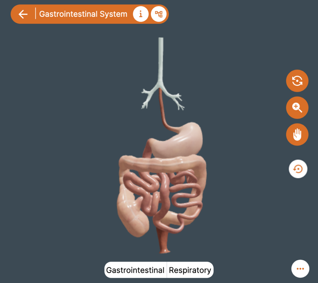
3D Model Embedded in the Greco-Roman Anatomical Atlas Copyright: ATLOMY (ERC StG GA 852550) Hebrew University of Jerusalem – Step into the Atlas and use it to explore the model and to learn about Aristotle’s ideas of the anatomy of the digestive system and about his anatomical terminology. You might recognize some terms! You will see […]
The 3D Model of the Digestive System according to Aristotle
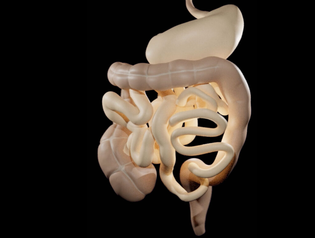
https://comparative-guts.net/wp-content/uploads/2023/02/Atlomy-Exhibit-4_2.mp4 The 3D Model of the Digestive System according to Aristotle Copyright: ATLOMY (ERC StG GA 852550) Hebrew University of Jerusalem – This 3-D model of the stomach and the intestines presents ATLOMY’s interpretation of the ancient text. While it might not be evident at first sight, this model differs from contemporary models of the […]
3D Modelling of the Guts according to Aristotle
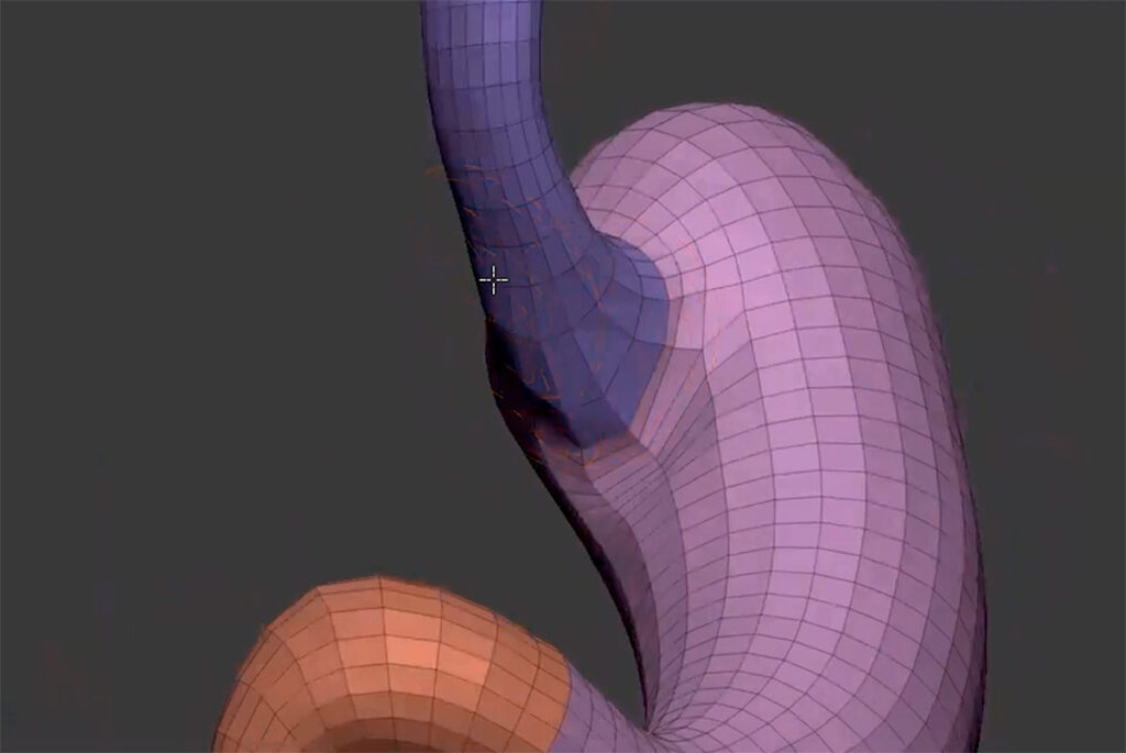
https://comparative-guts.net/wp-content/uploads/2023/02/Atlomy-Exhibit-3_1.mp4 3D Modelling of the Guts according to Aristotle Copyright: ATLOMY (ERC StG GA 852550) Hebrew University of Jerusalem – The next stage towards a visual reconstruction of Aristotle’s conception of guts is creating the 3D model. Based on the Table of Instructions the modellers use 3D-modelling software to create the model: from an initial […]
From Text to Model: The Guts according to Aristotle
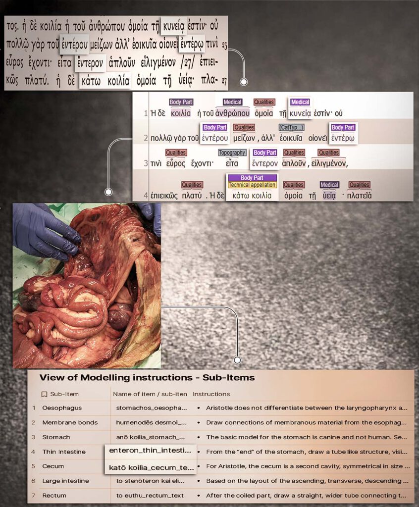
From Text to Model: The Guts according to Aristotle Copyright: ATLOMY (ERC StG GA 852550) Hebrew University of Jerusalem – A depiction of the first stages towards creating a 3D model of the human guts as described in Aristotle’s treatise Inquiries on Animals. The interdisciplinary team facilitates a unique analysis which bridges the gap between […]
From Observation to Text: The Guts according to Aristotle

From Observation to Text: The Guts according to Aristotle Copyright: ATLOMY (ERC StG GA 852550) Hebrew University of Jerusalem Bronze retractor, Roman, 1-100 CE. Science Museum, London. Attribution 4.0. International (CC BY 4.0) http://www.internetculturale.it/ – Sacrificial rituals were a common means to observe and learn about internal anatomy in ancient Greece. Aristotle investigated the body […]
Blasengeschwülste, Fig. 44-46: Ansichten eines Papilloms, Fig. 47-48: Ansichten eines Tumors (Kneise 1908: Tafel IX)
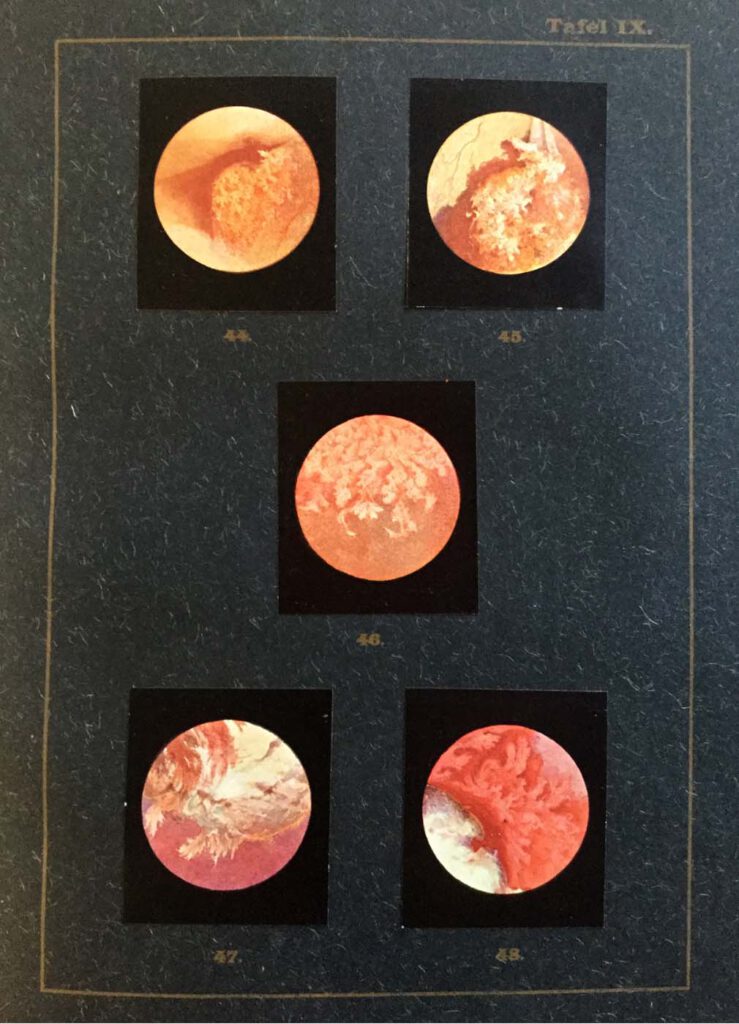
Blasengeschwülste, Fig. 44-46: Ansichten eines Papilloms, Fig. 47-48: Ansichten eines Tumors (Kneise 1908: Tafel IX) Kneise, Otto (1908): Handatlas der Cystskopie. Gebauer-Schwetschke: Halle a. Saale. — Attempts to access the interior of the body via orifices had already been made since antiquity, with particular attention being paid to the oral, genital, urinary and excretory organs. […]