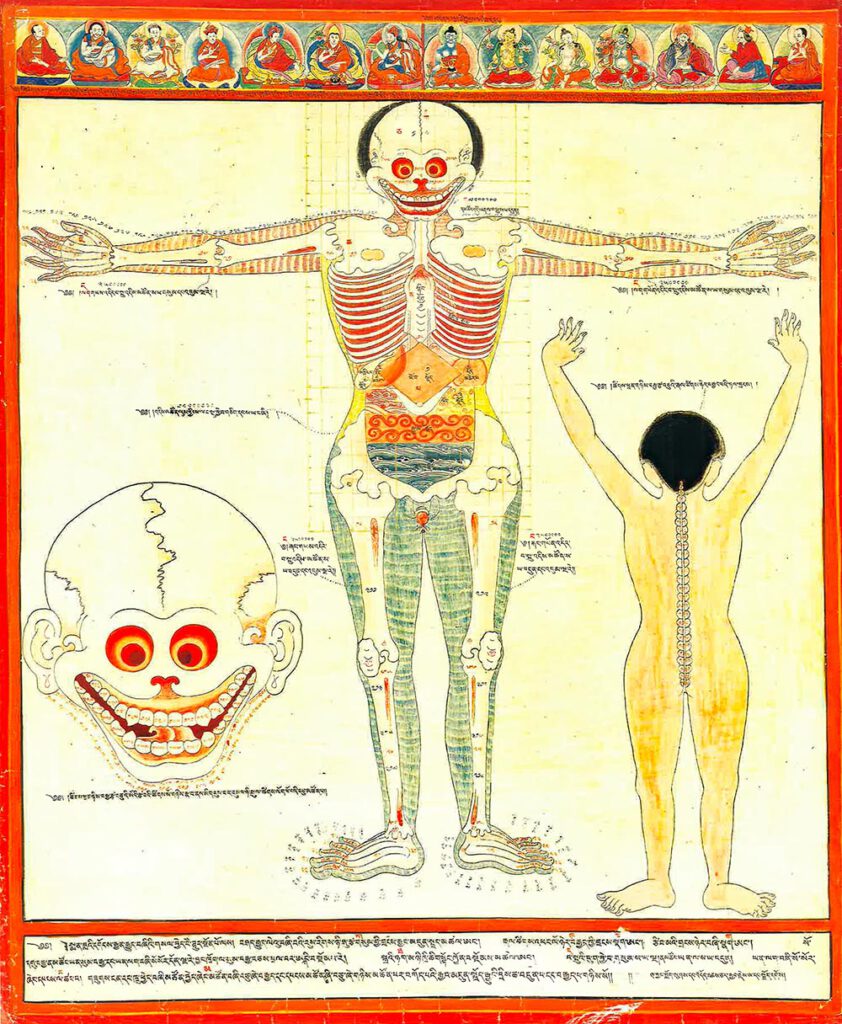
Major connecting blood vasculature of the body including gut with related vulnerable points
Major connecting blood vasculature of body including gut with related vulnerable points 20th century reproduction of 17th century CE painting. Held in Ulan-Ude, Republic of Buryatia, Russia. – Explicated in high detail in the traumatology section of the Four Tantras as developed from early military medical manuals, this painting depicts major blood vessels of the […]
Body metaphors including guts
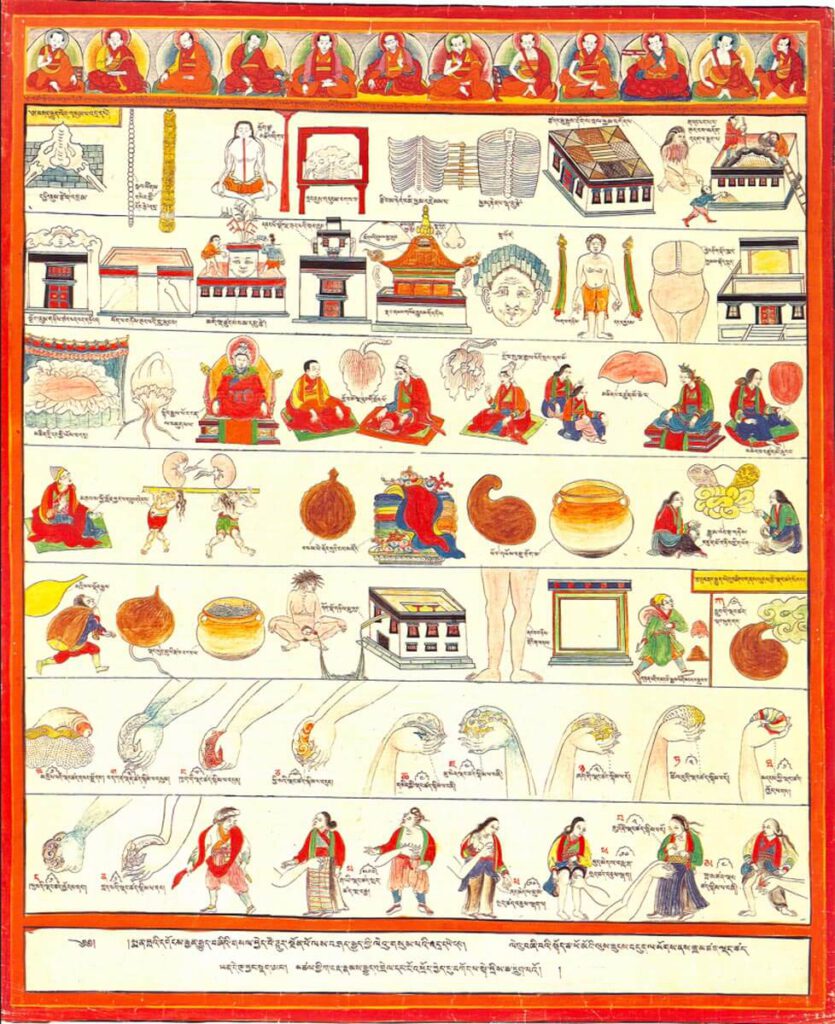
Body metaphors including guts 20th century reproduction of 17th century CE painting. Held in Ulan-Ude, Republic of Buryatia, Russia – The musculoskeletal system is likened to the architecture of a Tibetan temple, while internal organs, differentiated as solid (vital) and hollow (vessel) types, use different illustrative metaphors. The group of solid viscera include the heart, […]
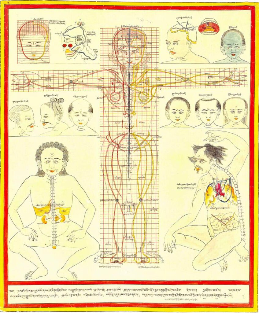
Location of internal organs on modular grid
Location of internal organs on modular grid 20th century reproduction of 17th century CE painting. Held in Ulan-Ude, Republic of Buryatia, Russia. – The central figures present the anterior and posterior views of the proper positions and proportions of anatomical structures from a theoretical perspective in relation to the spine by means of the Tibetan […]
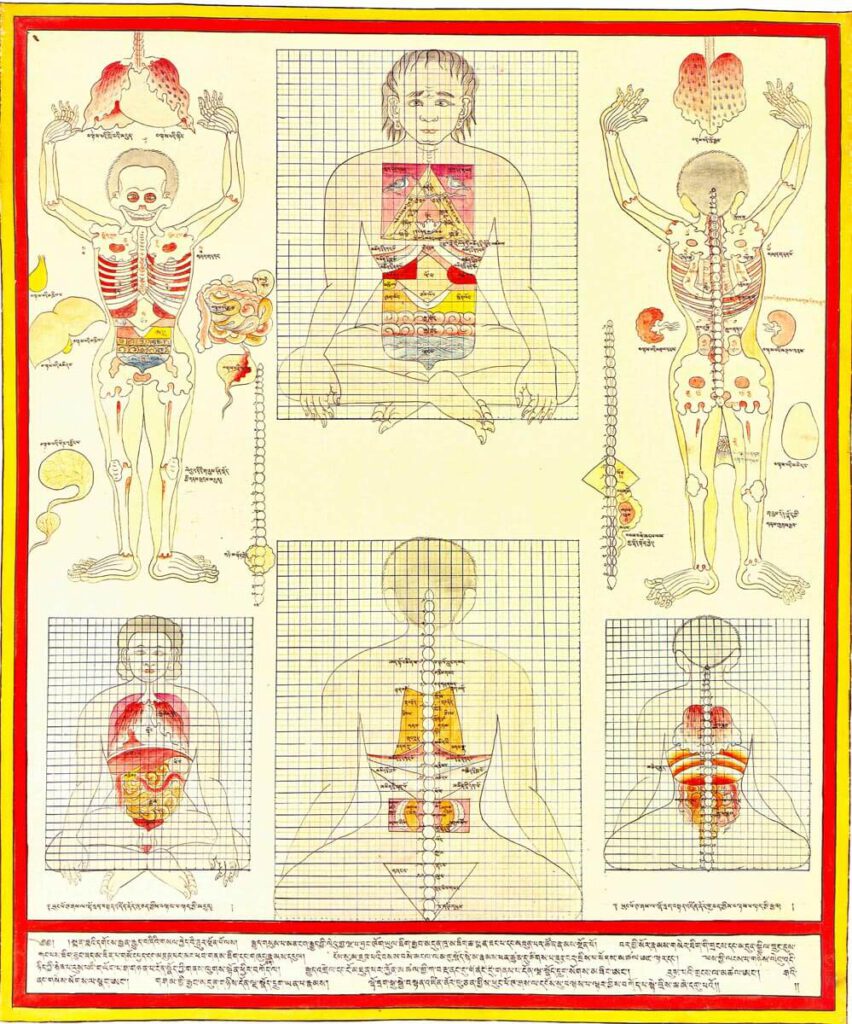
Anatomy of the gut, anterior view
Anatomy of the gut, anterior view 20th century reproduction of 17th century CE painting. Held in Ulan-Ude, Republic of Buryatia, Russia. – This painting displays an enumeration of body part components (e.g., bones, vertebrae, joints, etc.) and diagrams the proper associated relational measures in anterior view using a quadrant grid. The abdominal quadrant covers 12×8 […]
Nkisi power figure: “Mavungu”
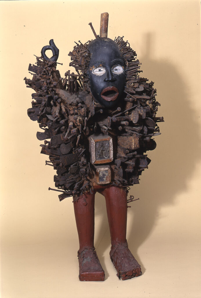
Nkisi power figure: ‘Mavungu’ Held in the Pitt Rivers Museum, Oxford. 1900.39.70. Figure carved of wood and painted, studded with iron nails and implements; a ‘power figure’. [JC 27/9/2001] – Kongo nkisi: nkisi are controversially known as “nail fetishes” and famously trouble the Western concept of “animism” and African religion. The concept of the fetish […]
Indian anatomical painting
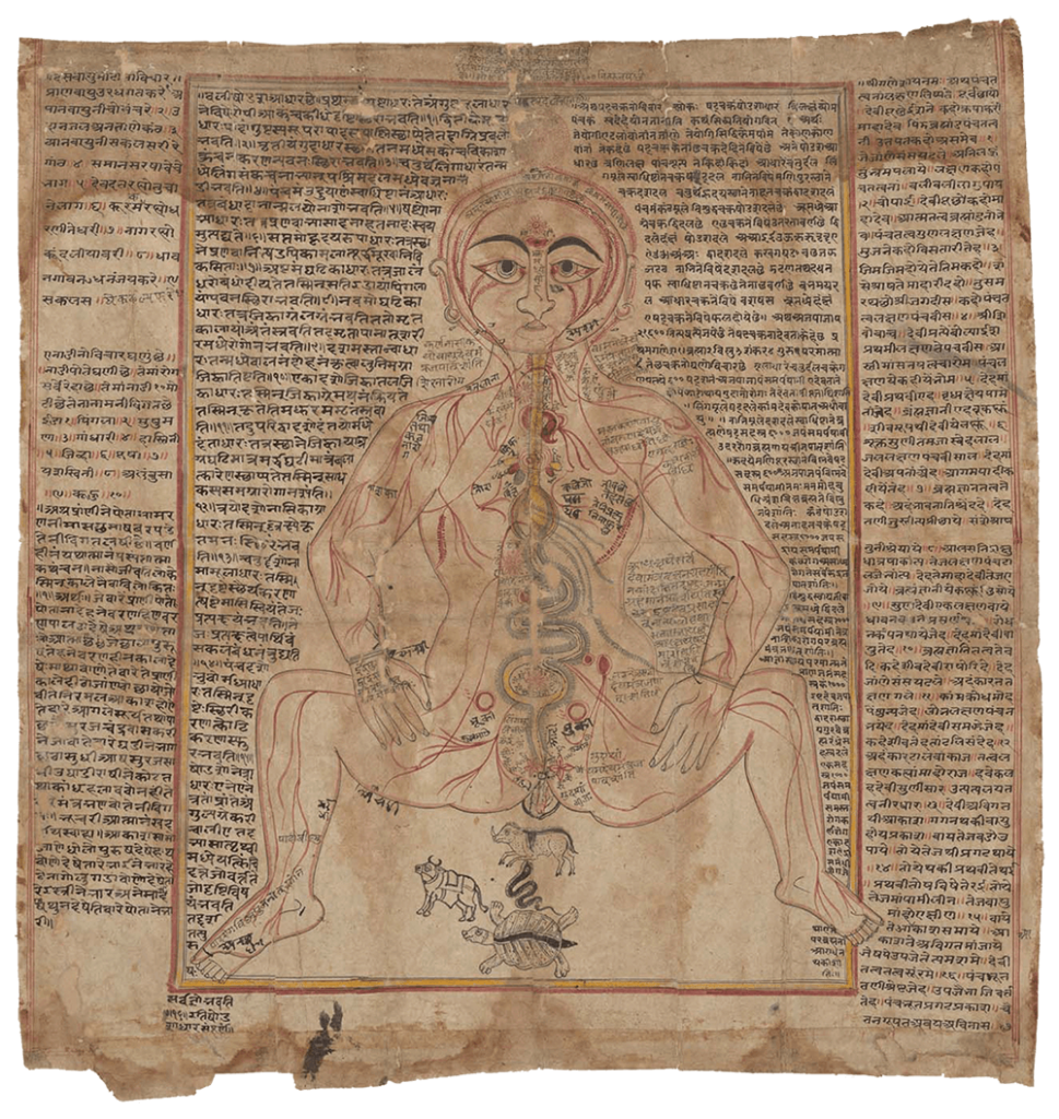
Indian anatomical painting Circa eighteenth century, western India. An old-Gujarati manuscript (circa 1900?). (Wellcome MS Indic d 74. Photo Wellcome Library, London.) Source: Wellcome Collection CC BY-NC-SA 4.0 – Premodern anatomical images in India were influenced by Persian drawings on anatomy, particularly during the Mughal dynasty’s reign in north India from the sixteenth to eighteenth […]
A human anatomical figure
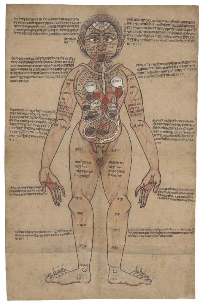
A human anatomical figure Drawing, Nepalese, ca. 1800 (?) Source: Wellcome Library no. 574912iWellcome Collection. Public Domain Mark. – Although India has thousands of manuscripts of premodern medical texts, only a handful contain anatomical illustrations. Text was crucially important for transmitting medical teachings. Figure 1 combines text with an illustration of the body and its […]
Towing bladder/hunting float.
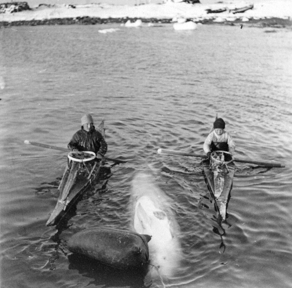
Towing bladder / hunting float keeping a hunted whale floating. Photo: Bodil Begtrup, Danish Arctic Institute – This important hunting implement, when hunting seals from a kayak is, despite of the name, usually made from the stomach of a narwhal or, if this is not available, from waterproof seal skin. Its length is usually 35-50 cm. […]
Diseases of the Stomach (cancer)
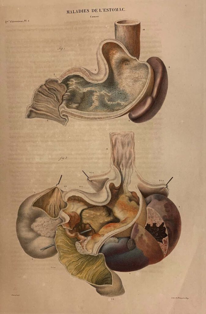
Diseases of the Stomach (cancer) lithograph printed by Benard and Frey after Antoine Touissant de Chazal (French, 1793-1854). From Jean Cruveilhier, Anatomie pathologique du corps humain (Paris: J. B. Baillière, 1829-1842), vol. 2, part 27, pl. 1. Huntington Library, Art Museum and Gardens, San Marino, California, RB 632011. – By the nineteenth century, publishing technology […]
Abdominal Dissection
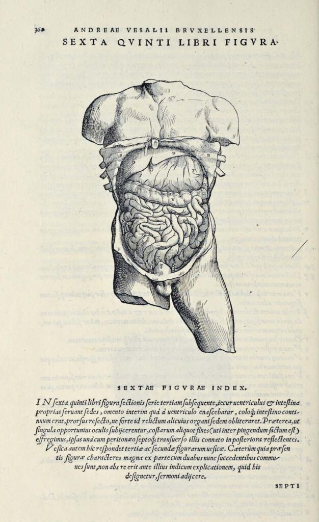
Abdominal Dissection woodcut after Jan Steven van Calcar (North Netherlandish, ca. 1515– ca. 1546). From Andreas Vesalius, De humani corporis fabrica libri septem (Basel: J. Oporinus, 1543), bk. 5, p. 360 [460], fig. 6. Getty Research Institute, Los Angeles, 84-B27611 – Ribs have been broken and the skin peeled back to display the liver, stomach, […]