Blasengeschwülste, Fig. 44-46: Ansichten eines Papilloms, Fig. 47-48: Ansichten eines Tumors (Kneise 1908: Tafel IX)
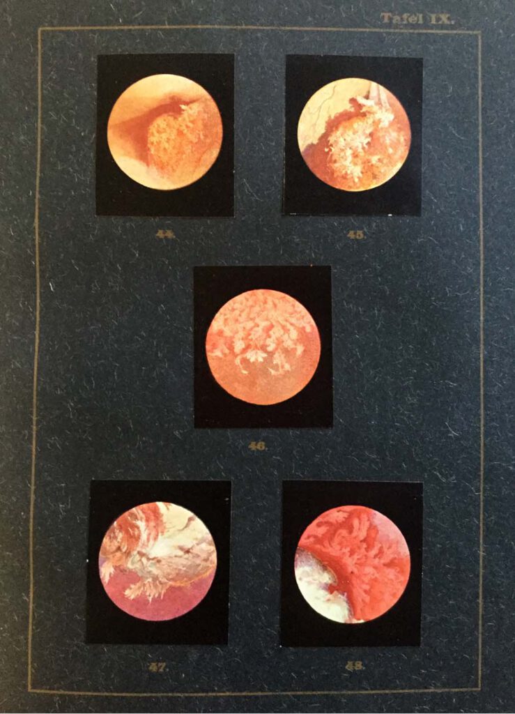
Blasengeschwülste, Fig. 44-46: Ansichten eines Papilloms, Fig. 47-48: Ansichten eines Tumors (Kneise 1908: Tafel IX) Kneise, Otto (1908): Handatlas der Cystskopie. Gebauer-Schwetschke: Halle a. Saale. — Attempts to access the interior of the body via orifices had already been made since antiquity, with particular attention being paid to the oral, genital, urinary and excretory organs. […]
Lehrbuch der urologischen Diagnostik
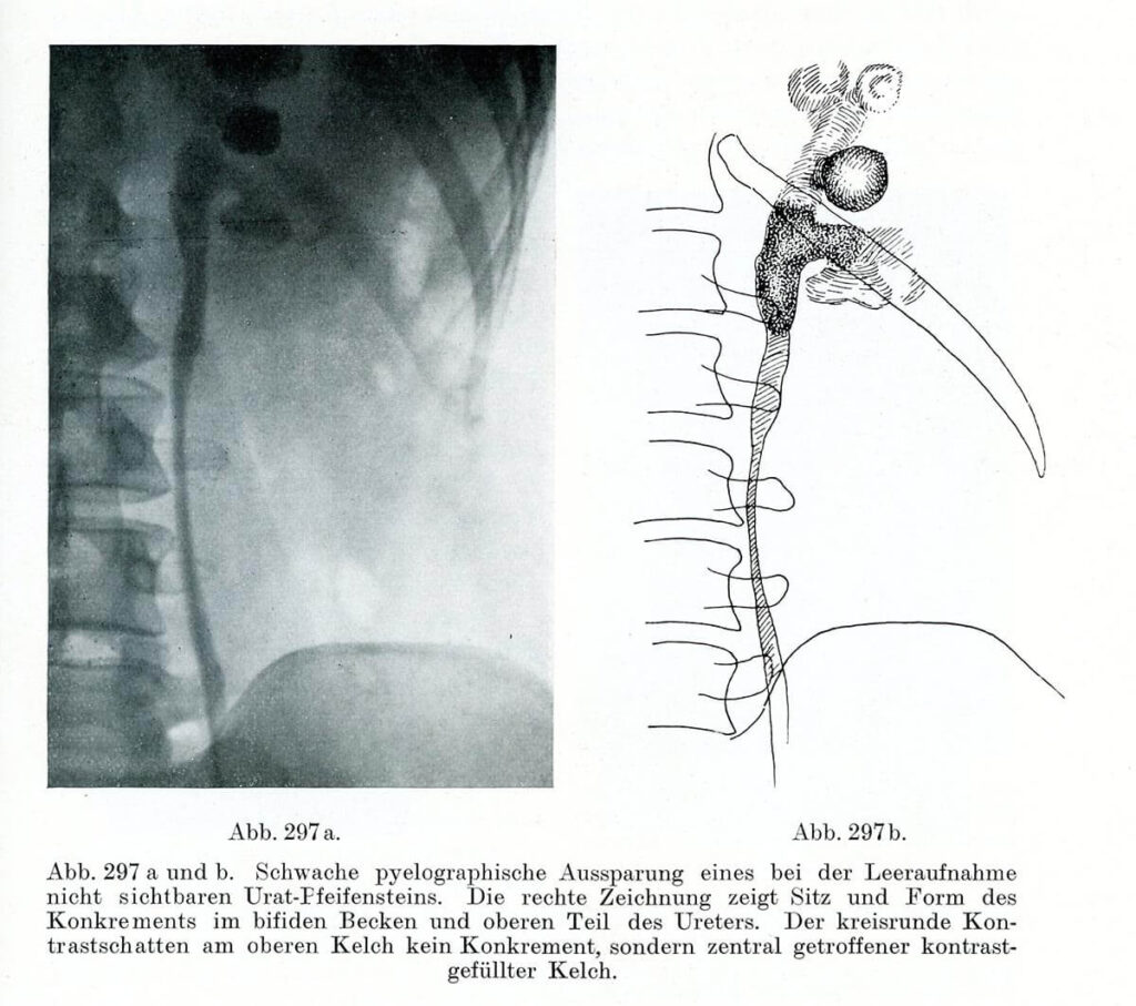
Pyeolographische Darstellung eines Konkrements im Ureter Leopold Casper und Edwin Picard (1930) Lehrbuch der urologischen Diagnostik. Thieme, Leipzig, S. 397 – Right after the discovery of X-rays, in the late 19th century, innumerable attempts were made to make this novel technique useful for medicine. While the radiological detection of bones quickly became a routine procedure, […]
Carcinoma colloides peritonei

Carcinoma colloides peritonei From: Emil Ponfick: Topographischer Atlas der Medizinisch-Chirurgischen Diagnostik / Topographic atlas of medico-surgical diagnosis, G. Fischer. Jena 1900-1905 Artist: Dr. Emil Löschmann, Breslau — The attempts of the German pathologist Emil Ponfick, who in 1901 attempted to reduce the gap between drawing and original object, show how objectives and implementation should be brought together […]
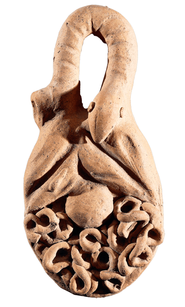
A female ‘open torso’ figurine

A female ‘open torso’ figurine Nottingham Castle Museum – This female figurine was discovered at the sanctuary of the Graeco-Roman goddess Artemis/Diana Nemorensis at Lake Nemi in Italy in 1885. It was excavated and recovered from a sacred pit where it had been ritually disposed of sometime in antiquity after it had ceased to be […]
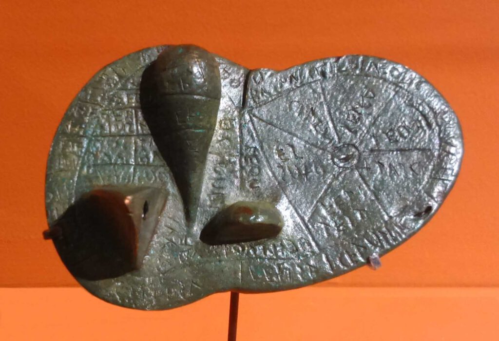
A polyvisceral plaque
A polyvisceral plaque Museo Nazionale Etrusco Di Villa Giulia – This Etruscan polyvisceral plaque from Tessennano in Latium in Italy is thought to date from around 400 BCE. Anatomical votives like these were deposited in sanctuaries and temples as offerings to the gods, generally interpreted as a gesture of gratitude for some manner of divine […]
The ‘Piacenza Liver’
The ‘Piacenza Liver’ Municipale Museum of Piacenza CC0 1.0 Universal (CC0 1.0) – This Etruscan bronze life-size model of a sheep’s liver was discovered in a field near Gossolengo in Piacenza in Italy in 1877 and is thought to date from the late second or early first century BCE. Although its precise purpose is not […]
Athenian red-figure cup attributed to Oltos, examination of the liver of a sacrificial victim
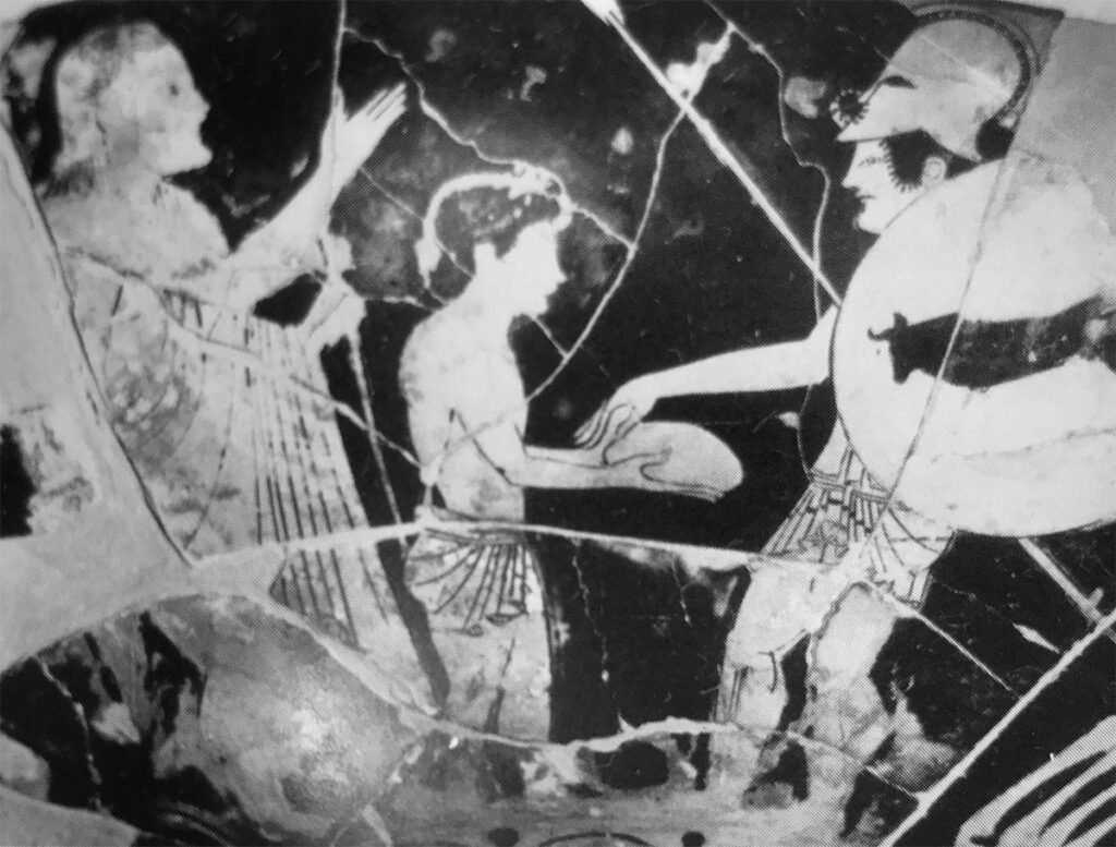
Athenian red-figure cup attributed to Oltos examination of the liver of a sacrificial victim (‘hieroscopy’), c.500 B.C.Cervetri, Museo Nazionale Cerite Photo: Jaime Ardiles-Arce – Representation of warfare is a feature of Greek painted pottery from the earliest figurative images from the eighth century BCE. In the second half of the sixth century and into the […]
Anonymous, K. Sharḥ al-Maqāma al-Ṣalāḥiyya fī l-Khayl wa-al-Bayṭāra
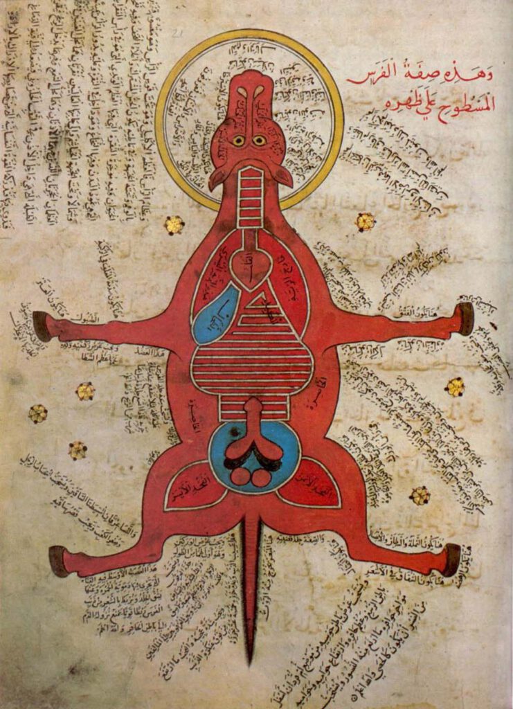
Anonymous, K. Sharḥ al-Maqāma al-Ṣalāḥiyya fī l-Khayl wa-al-Bayṭāra (Commentary on the Maqāma Ṣalāḥiyya on Horses and Venterinary) Istanbul University Library MS 4689 – Egypt, 15th c. – Hyppiatry has a long tradition in Islamic literature. Works dealing with horses may adopt different literary genres and cover a wide range of disciplines, from veterinary sciences to […]
Representation of a woman with the distribution of the blood vessels and the internal organs (f. 15v)
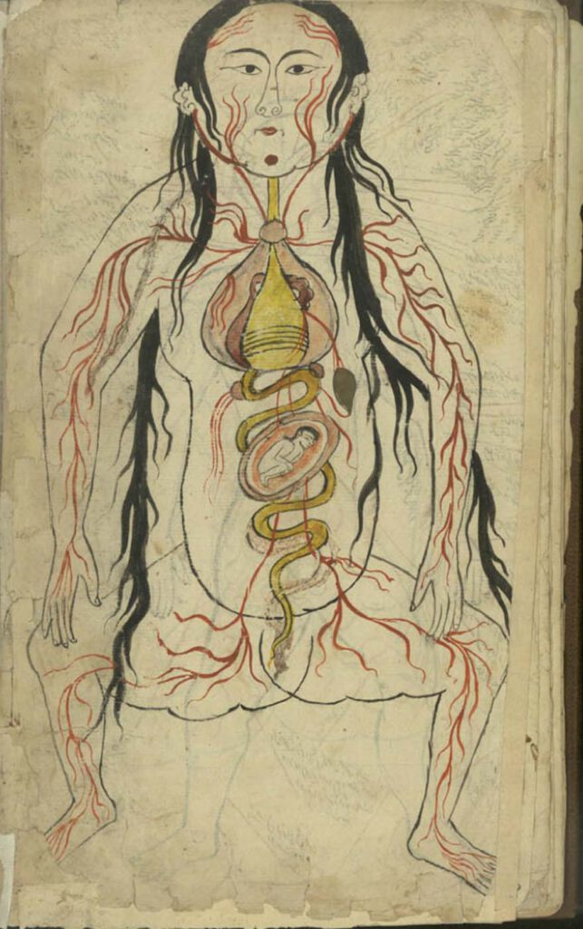
Representation of a woman with the distribution of the blood vessels and the internal organs (f. 15v) Manṣūr ibn Ilyās (fl. 14th c.), Tashrīḥ-i badan-i insān. Images from: Teheran MS Majlis 7430. Undated. This image represents the heart, lungs, liver, esophagus/larynx, stomach, intestines, kidneys, and spleen). As in the male representation, the organs involved in […]
Representation of a man with the distribution of the blood vessels and the internal organs (f. 13v)
Representation of a man with the distribution of the blood vessels and the internal organs (f. 13v) Manṣūr ibn Ilyās (fl. 14th c.), Tashrīḥ-i badan-i insān. Images from: Teheran MS Majlis 7430. Undated. The illustration depicts the heart, lungs, liver, esophagus/larynx, stomach, intestines, kidneys, and spleen. The organs involved in digestion are coloured in yellow. […]