Brain Frog
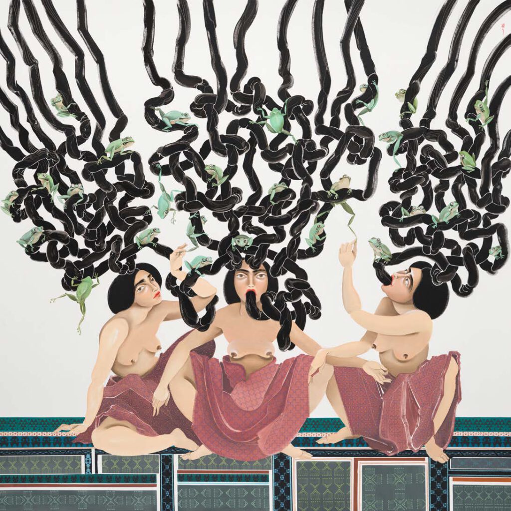
Brain Frog Hayv Kahraman Brain Frog, 2022 Oil on panel 203.2 × 203.2 cm 80 × 80 in (KAHR 2022001) Read the History
3D Model Embedded in the Greco-Roman Anatomical Atlas
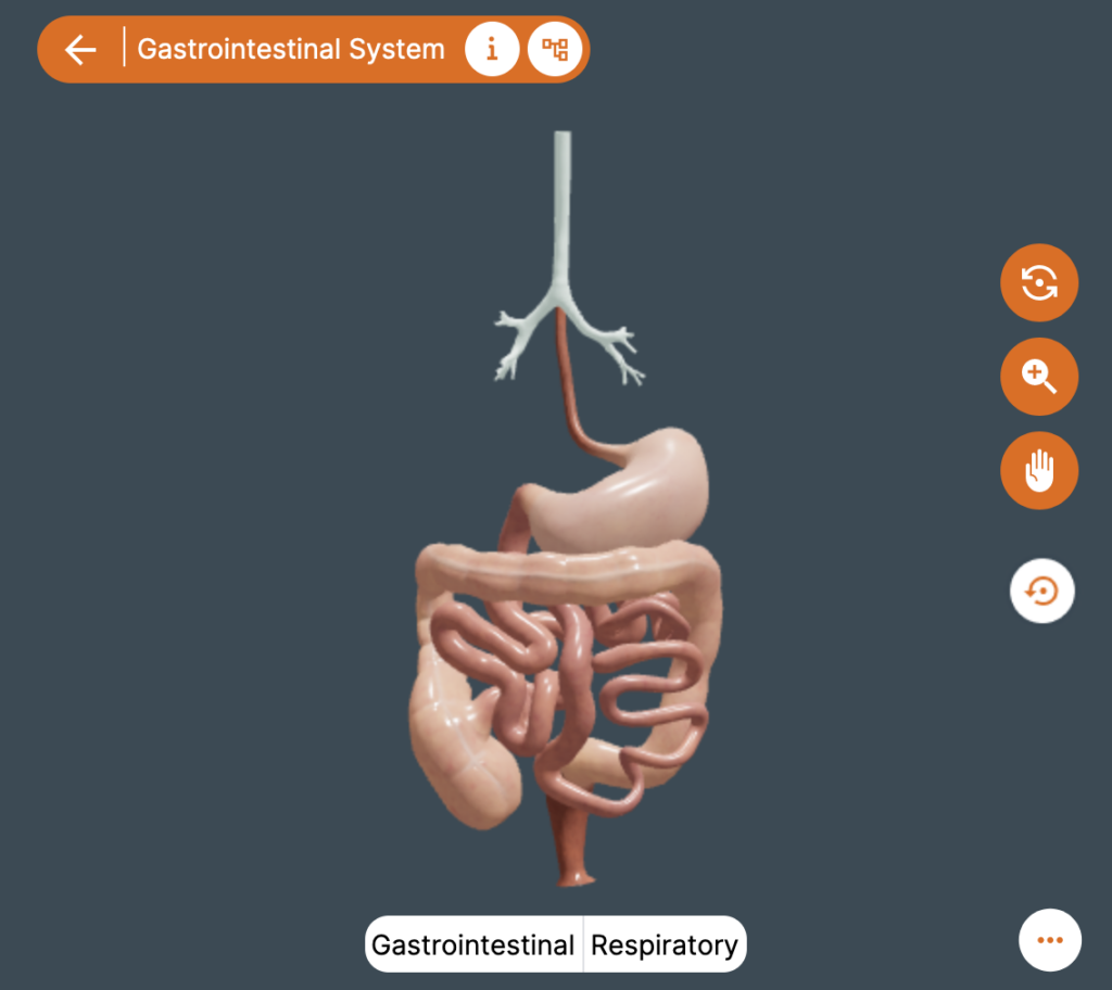
3D Model Embedded in the Greco-Roman Anatomical Atlas Copyright: ATLOMY (ERC StG GA 852550) Hebrew University of Jerusalem – Step into the Atlas and use it to explore the model and to learn about Aristotle’s ideas of the anatomy of the digestive system and about his anatomical terminology. You might recognize some terms! You will see […]
The 3D Model of the Digestive System according to Aristotle
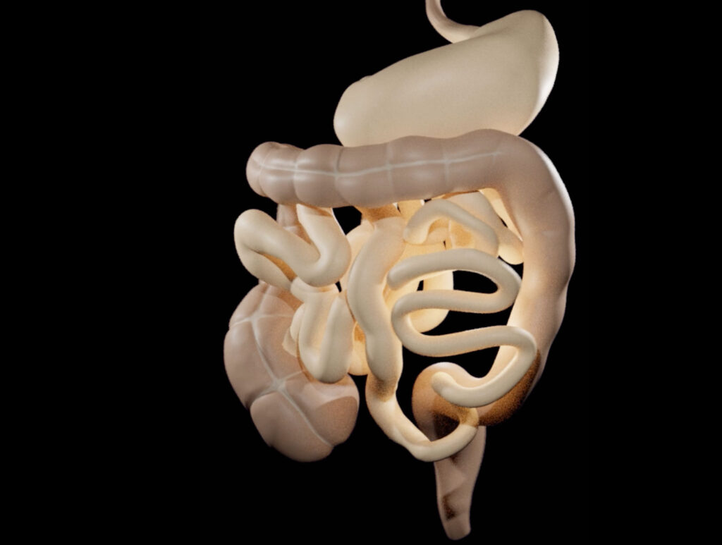
https://comparative-guts.net/wp-content/uploads/2023/02/Atlomy-Exhibit-4_2.mp4 The 3D Model of the Digestive System according to Aristotle Copyright: ATLOMY (ERC StG GA 852550) Hebrew University of Jerusalem – This 3-D model of the stomach and the intestines presents ATLOMY’s interpretation of the ancient text. While it might not be evident at first sight, this model differs from contemporary models of the […]
3D Modelling of the Guts according to Aristotle
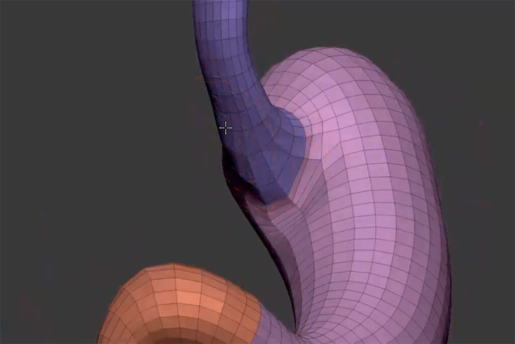
https://comparative-guts.net/wp-content/uploads/2023/02/Atlomy-Exhibit-3_1.mp4 3D Modelling of the Guts according to Aristotle Copyright: ATLOMY (ERC StG GA 852550) Hebrew University of Jerusalem – The next stage towards a visual reconstruction of Aristotle’s conception of guts is creating the 3D model. Based on the Table of Instructions the modellers use 3D-modelling software to create the model: from an initial […]
From Text to Model: The Guts according to Aristotle
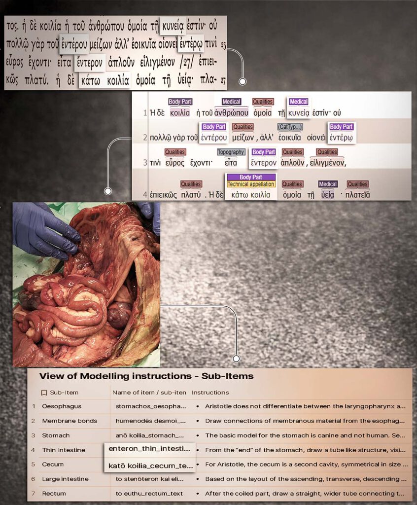
From Text to Model: The Guts according to Aristotle Copyright: ATLOMY (ERC StG GA 852550) Hebrew University of Jerusalem – A depiction of the first stages towards creating a 3D model of the human guts as described in Aristotle’s treatise Inquiries on Animals. The interdisciplinary team facilitates a unique analysis which bridges the gap between […]
From Observation to Text: The Guts according to Aristotle

From Observation to Text: The Guts according to Aristotle Copyright: ATLOMY (ERC StG GA 852550) Hebrew University of Jerusalem Bronze retractor, Roman, 1-100 CE. Science Museum, London. Attribution 4.0. International (CC BY 4.0) http://www.internetculturale.it/ – Sacrificial rituals were a common means to observe and learn about internal anatomy in ancient Greece. Aristotle investigated the body […]
Blasengeschwülste, Fig. 44-46: Ansichten eines Papilloms, Fig. 47-48: Ansichten eines Tumors (Kneise 1908: Tafel IX)
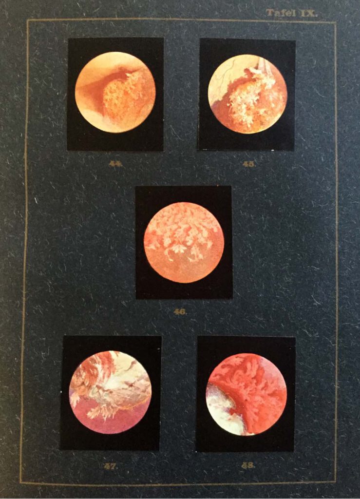
Blasengeschwülste, Fig. 44-46: Ansichten eines Papilloms, Fig. 47-48: Ansichten eines Tumors (Kneise 1908: Tafel IX) Kneise, Otto (1908): Handatlas der Cystskopie. Gebauer-Schwetschke: Halle a. Saale. — Attempts to access the interior of the body via orifices had already been made since antiquity, with particular attention being paid to the oral, genital, urinary and excretory organs. […]
Lehrbuch der urologischen Diagnostik
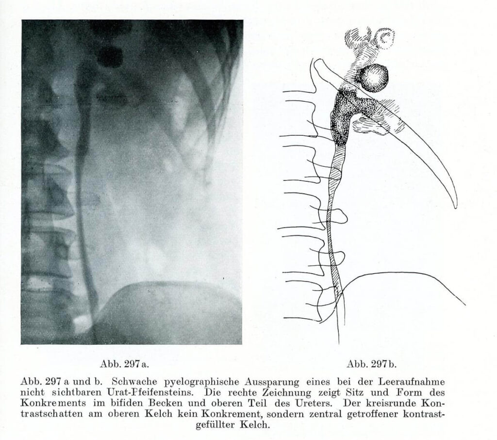
Pyeolographische Darstellung eines Konkrements im Ureter Leopold Casper und Edwin Picard (1930) Lehrbuch der urologischen Diagnostik. Thieme, Leipzig, S. 397 – Right after the discovery of X-rays, in the late 19th century, innumerable attempts were made to make this novel technique useful for medicine. While the radiological detection of bones quickly became a routine procedure, […]
Carcinoma colloides peritonei

Carcinoma colloides peritonei From: Emil Ponfick: Topographischer Atlas der Medizinisch-Chirurgischen Diagnostik / Topographic atlas of medico-surgical diagnosis, G. Fischer. Jena 1900-1905 Artist: Dr. Emil Löschmann, Breslau — The attempts of the German pathologist Emil Ponfick, who in 1901 attempted to reduce the gap between drawing and original object, show how objectives and implementation should be brought together […]