Skip to content
Comparative Guts
Comparative
Guts
Search
Search
Close this search box.
Menu
Solids
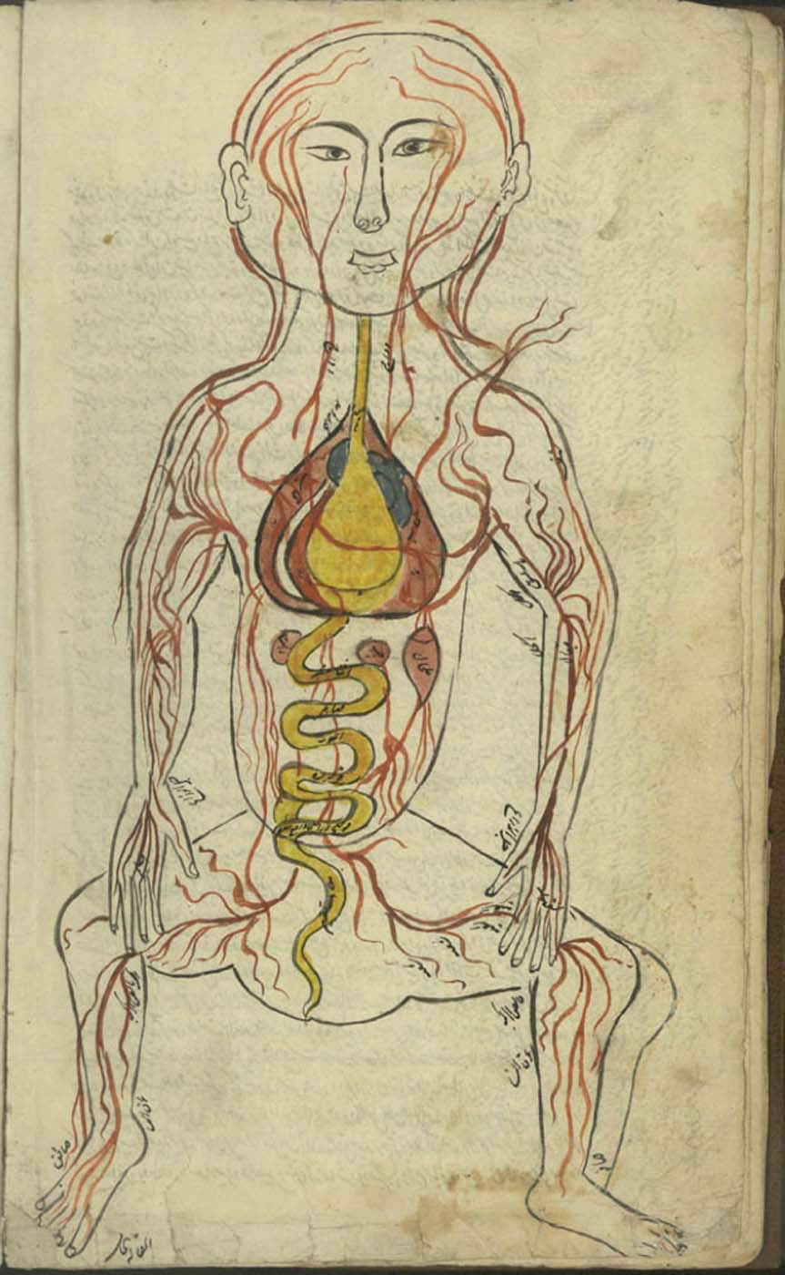
Guido da Vigevano’s Anathomia, Figure 8
Representation of a man with the distribution of the blood vessels and the internal organs (f. 13v)
Communication of the connectors of the five solid vessels with the heart.
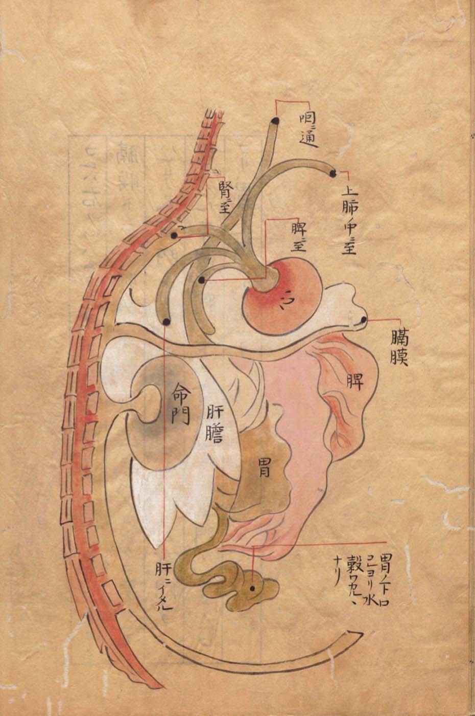
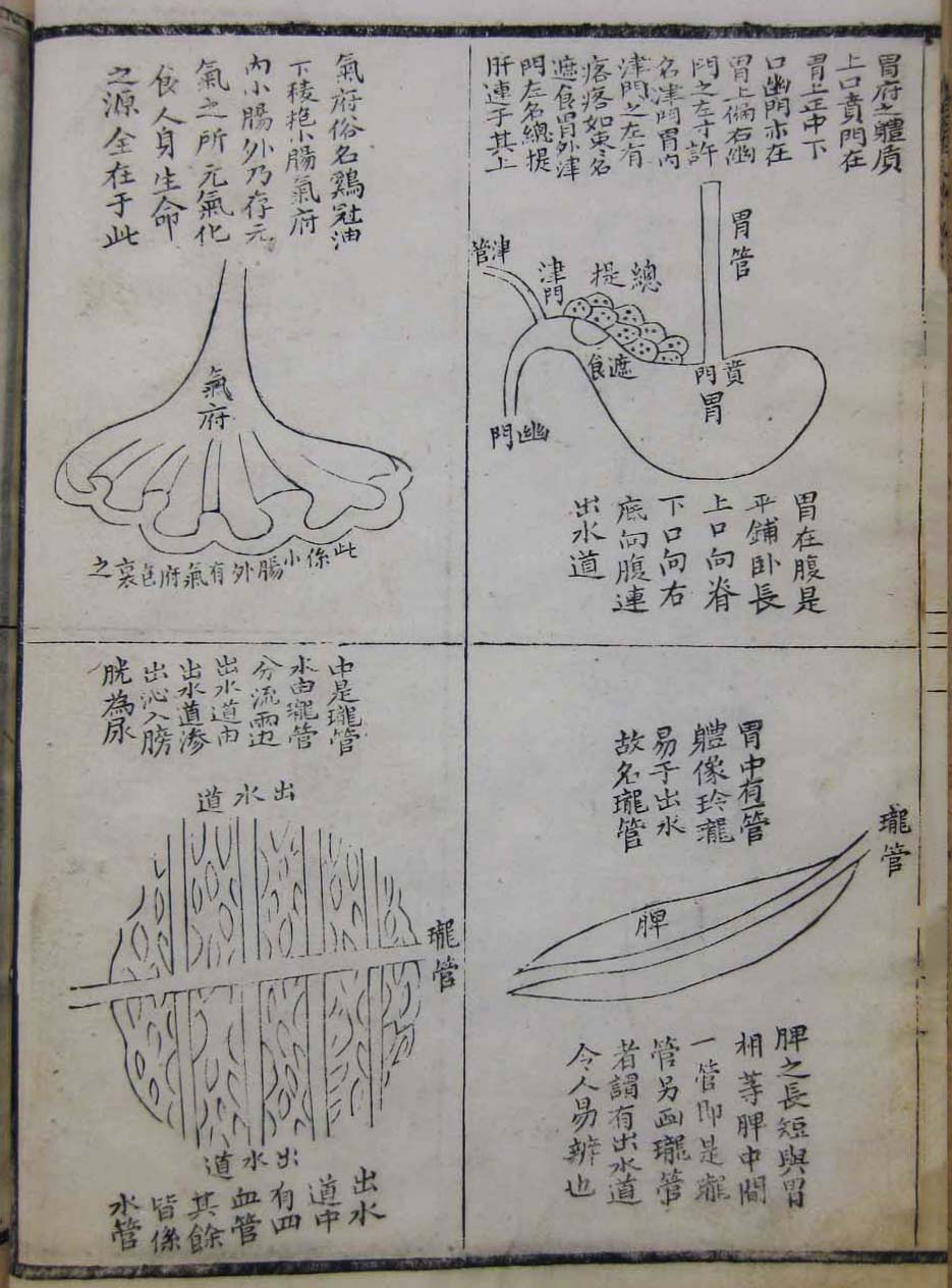
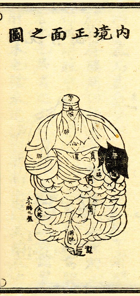
Manase Dōsan 曲直瀬道三, Hyakufuku zusetsu
Red-figured terracotta krater
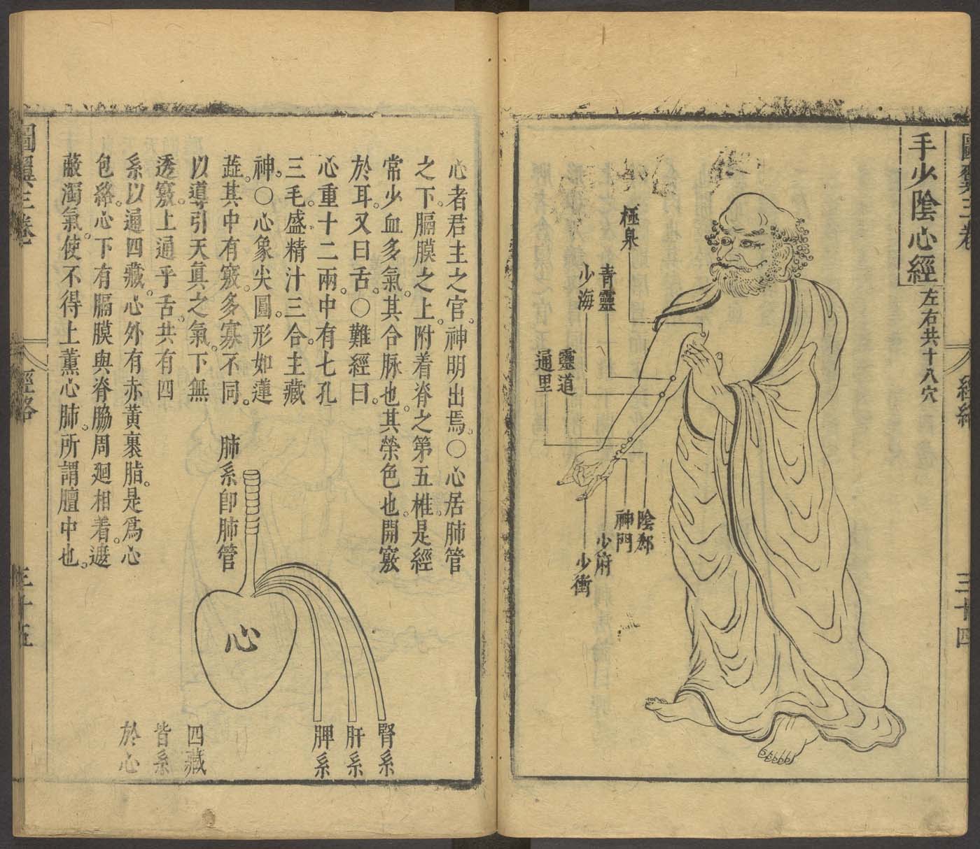
The hand lesser yin heart channel
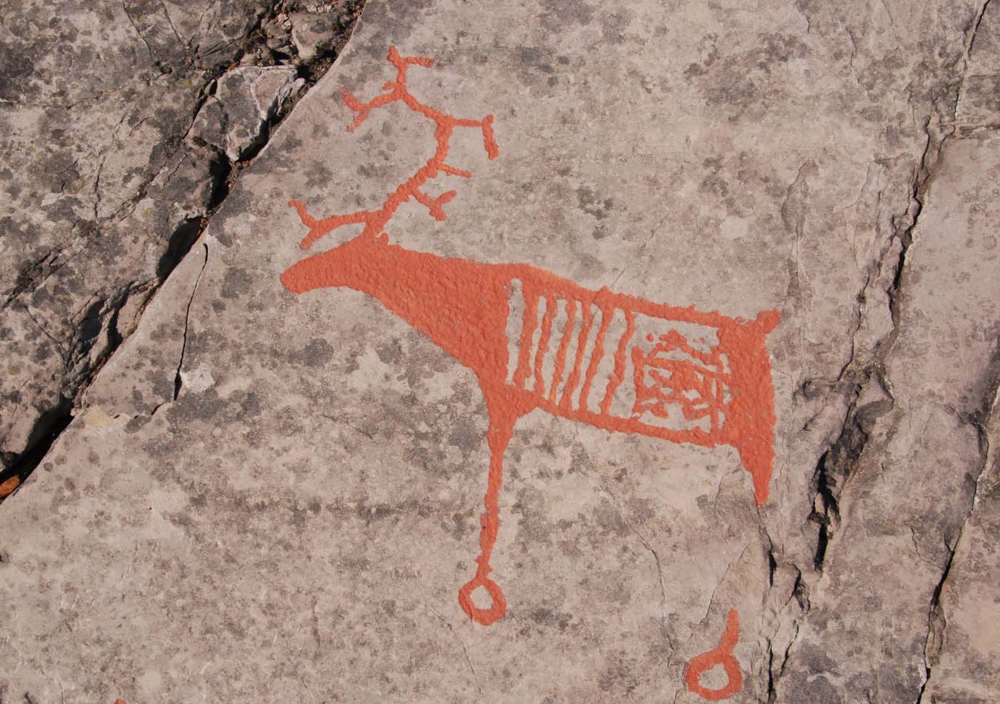
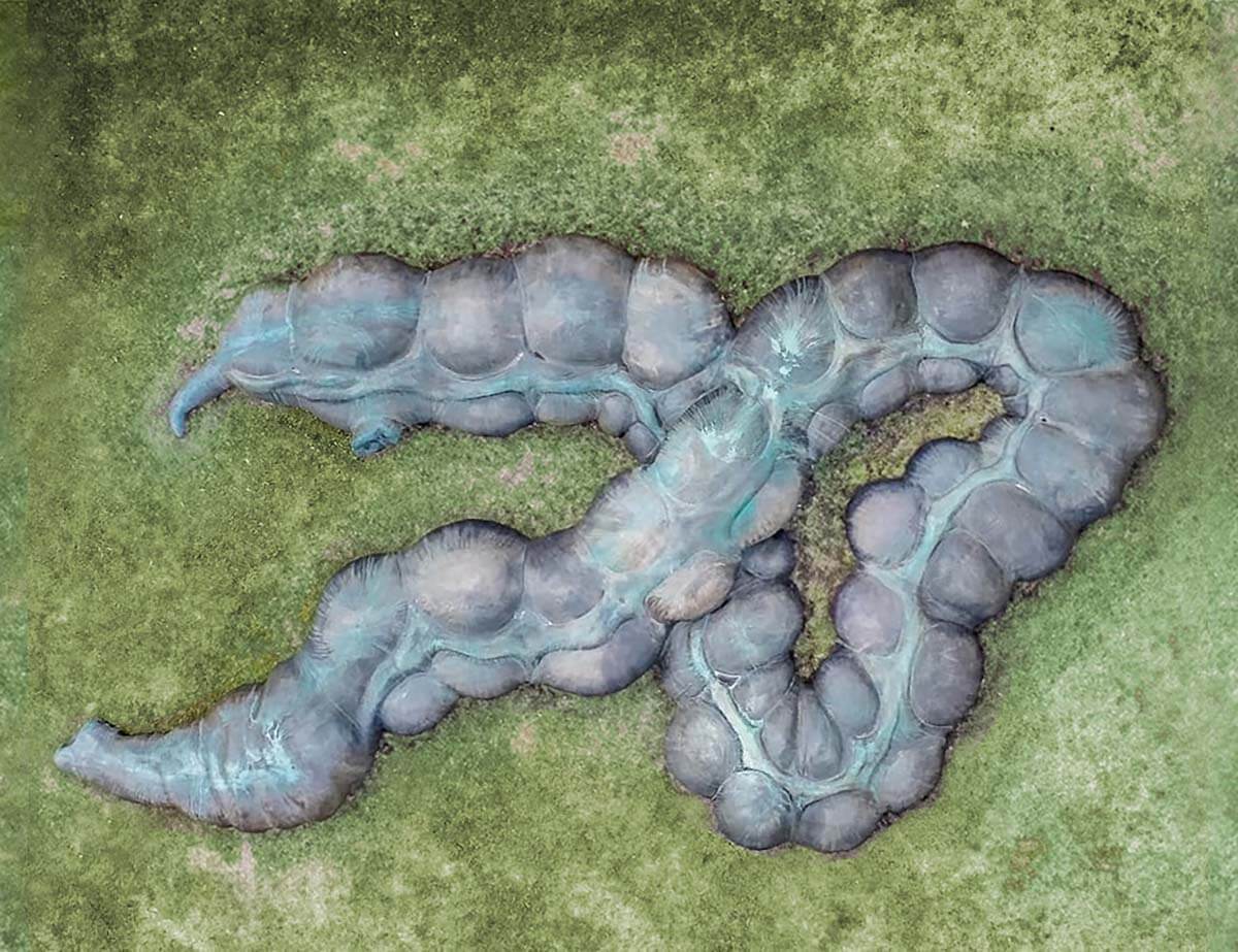
Rock carving of reindeer with intestines
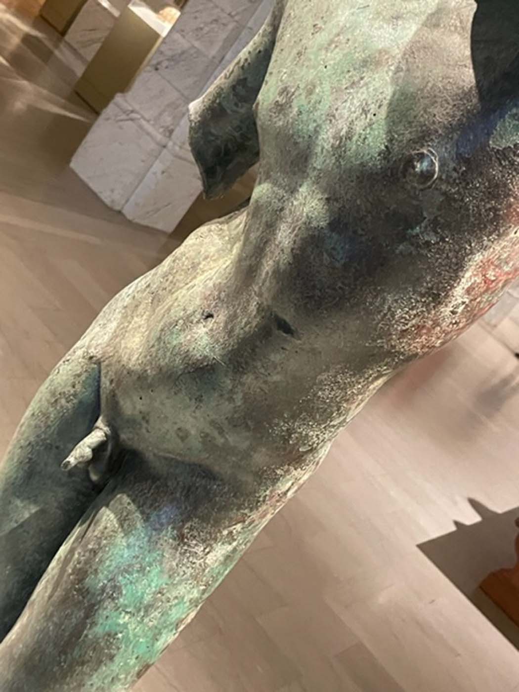
Apollo the Python-Slayer or Cleveland Apollo
Organs of the abdomen
3D Model Embedded in the Greco-Roman Anatomical Atlas
Anonymous,
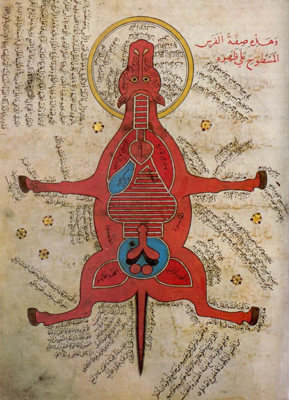
K. Sharḥ al-Maqāma al-Ṣalāḥiyya fī l-Khayl wa-al-Bayṭāra
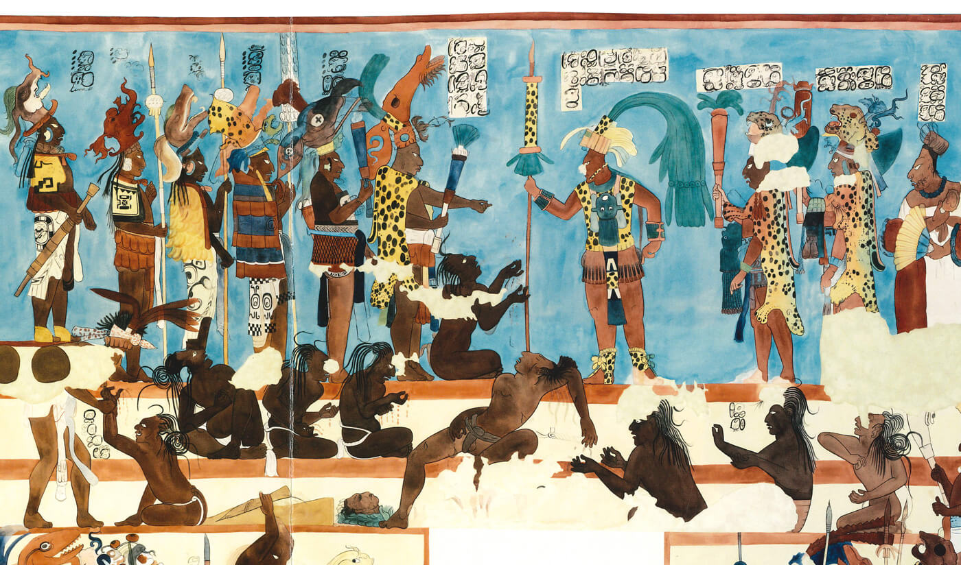
Battle scene and their aftermath
Red-figured terracotta bell-krater
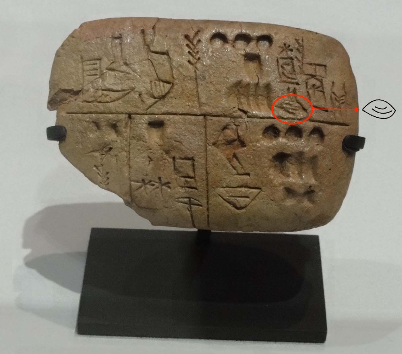
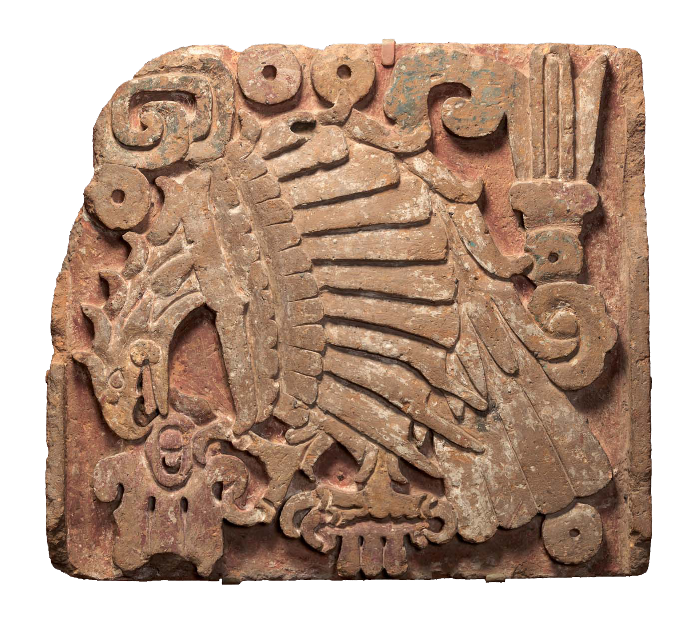
A polyvisceral plaque
ŠÀ, libbu
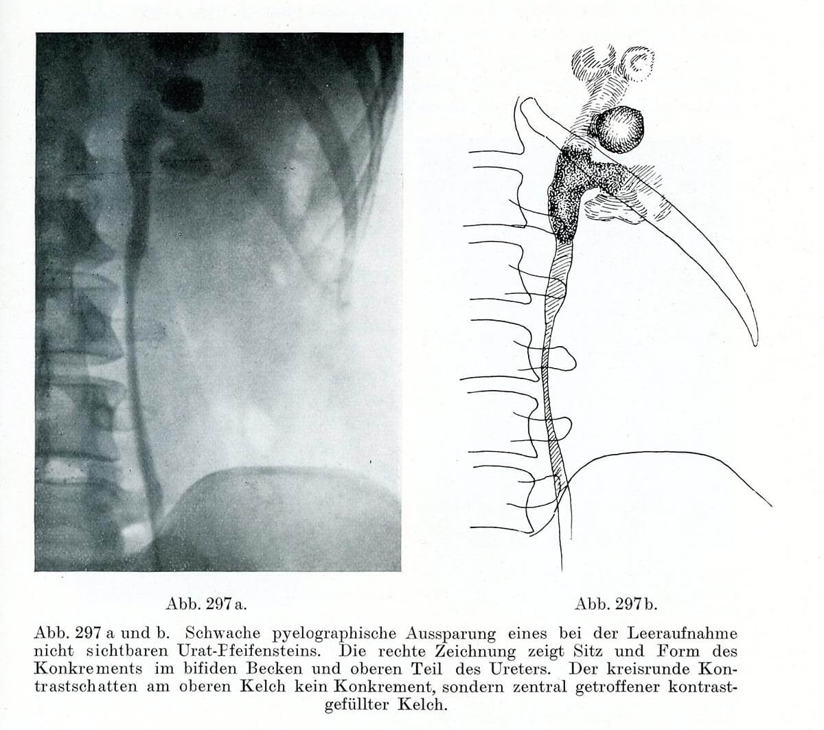
Lehrbuch der urologischen Diagnostik
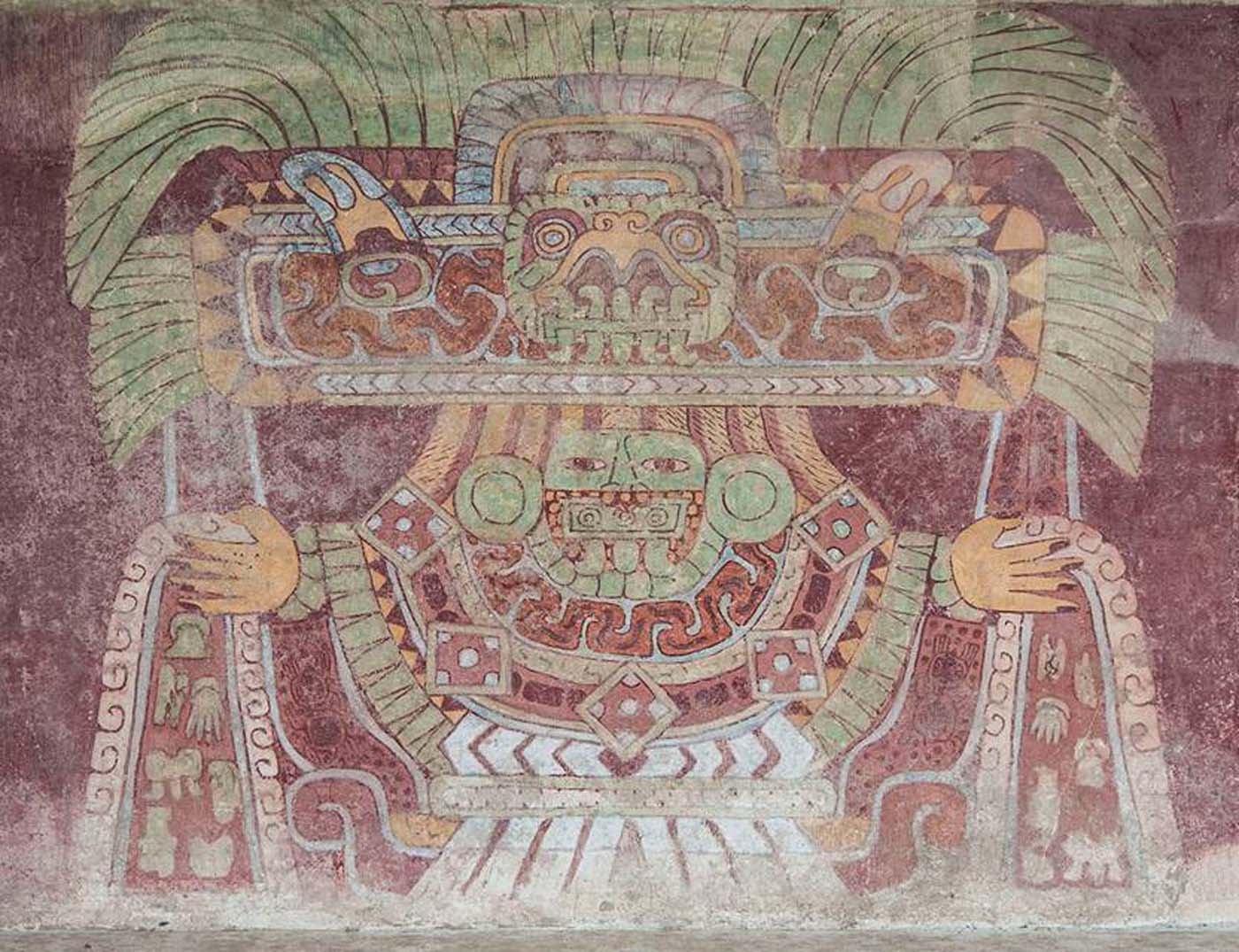
Depiction of a mortuary bundle at Teotihuacan
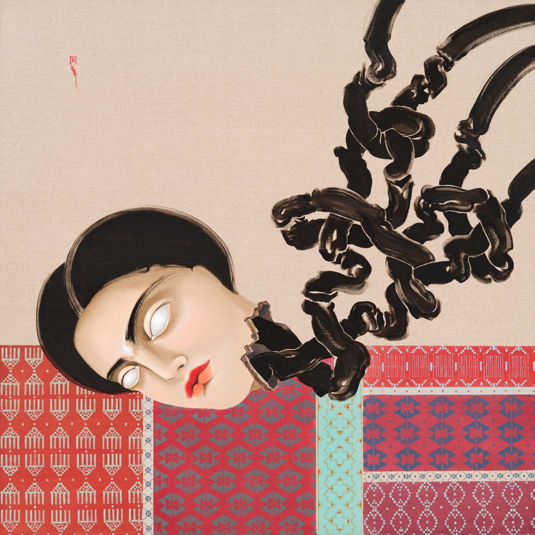
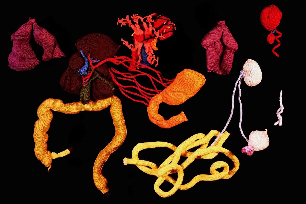
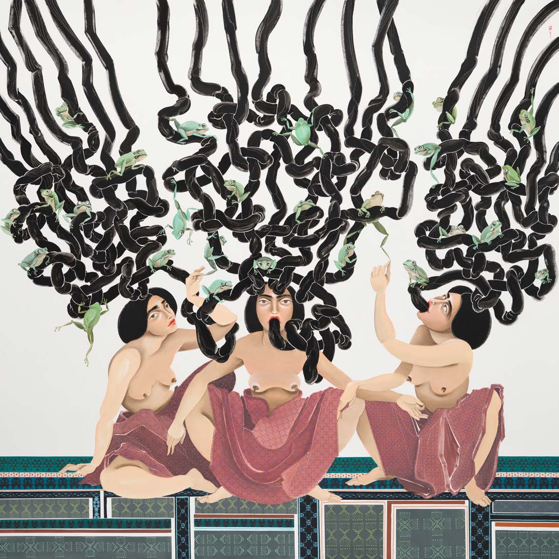
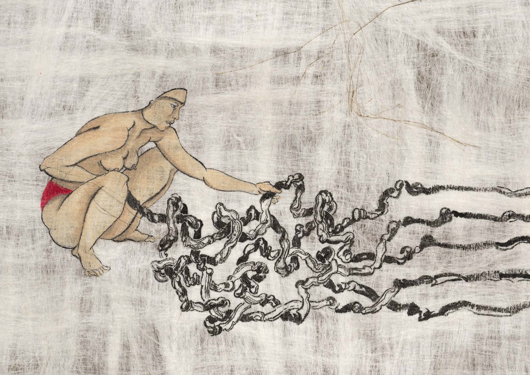
Entanglements with torshi no.2
Sugita Gempaku’s Kaitai shinsho
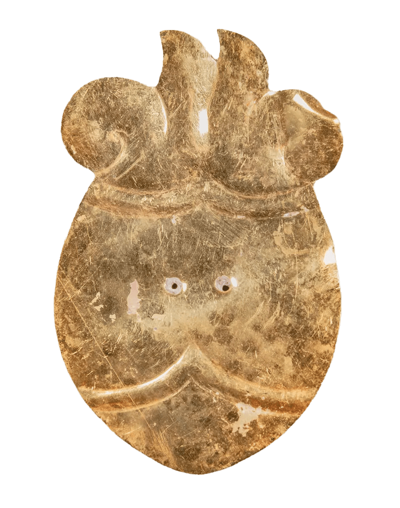
Stylized heart rendered in sheet gold
NeuroBust no.5
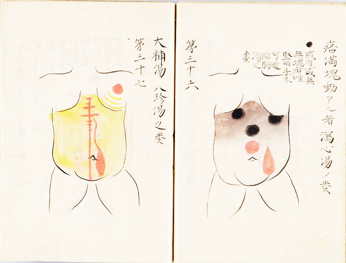
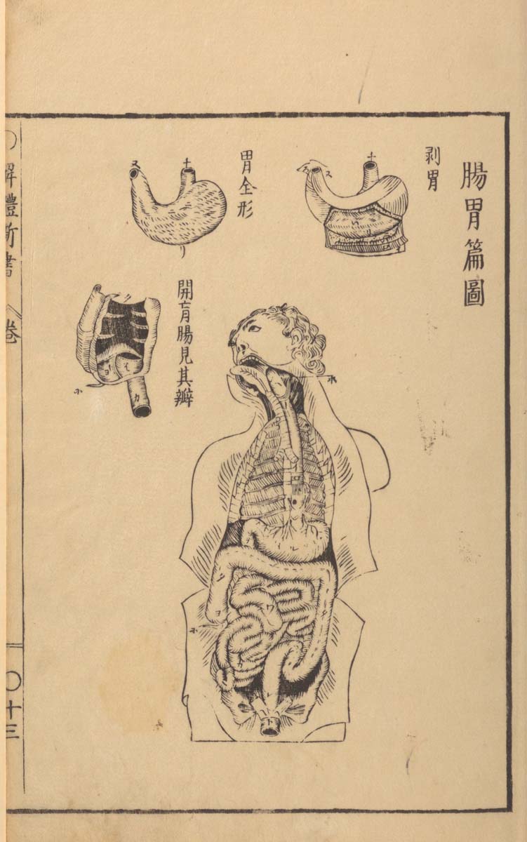
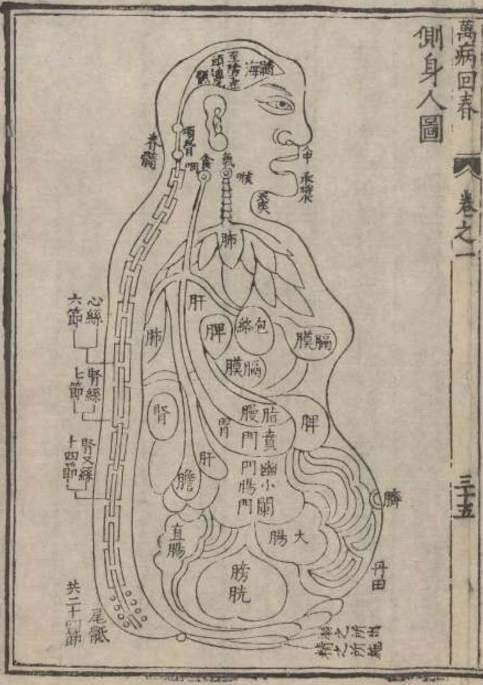
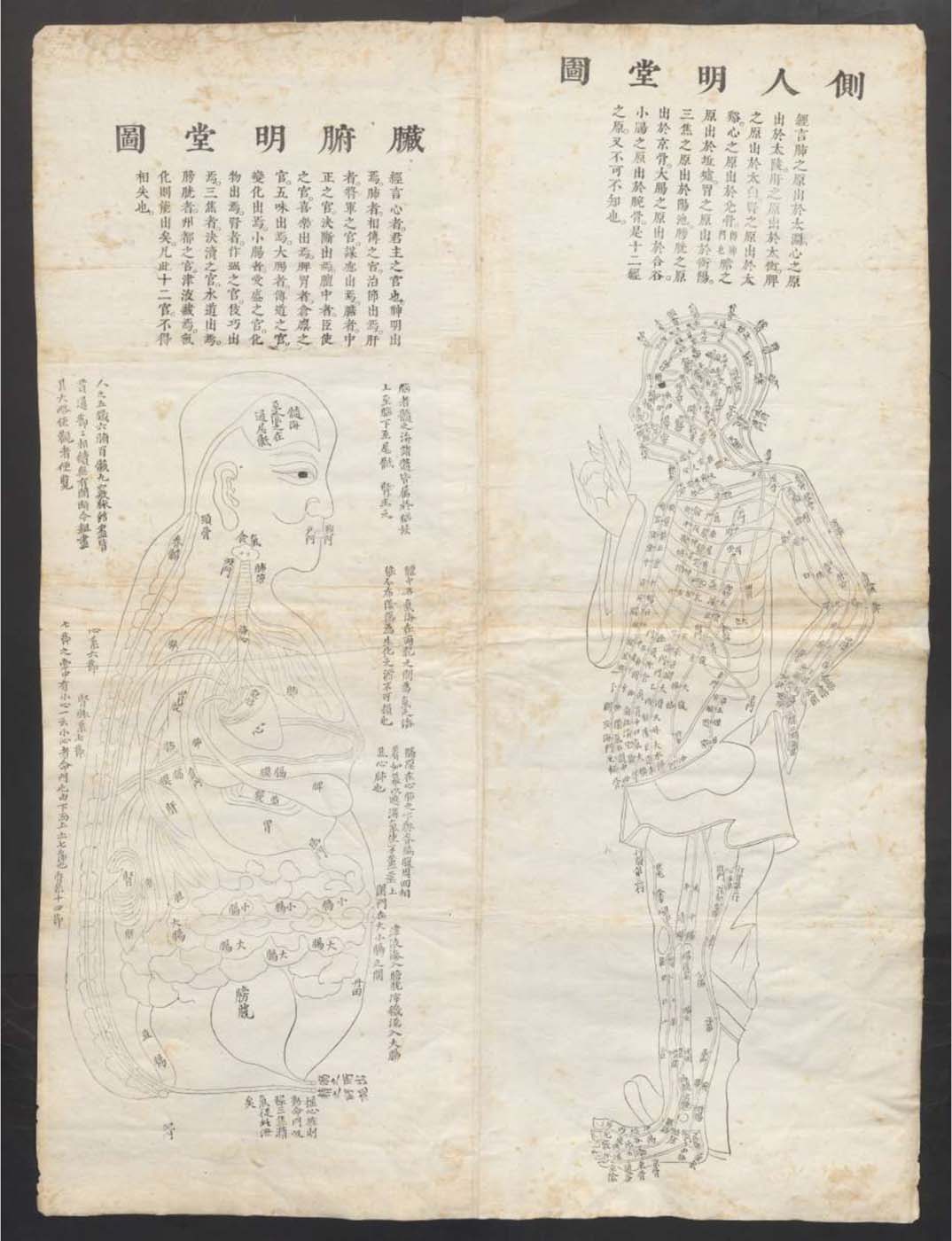
1597 reprint of Wanbing huichun “Diagram of Man’s Side-Body”
Mary Reid Kelley with Patrick Kelley,
This is Offal
(2015)
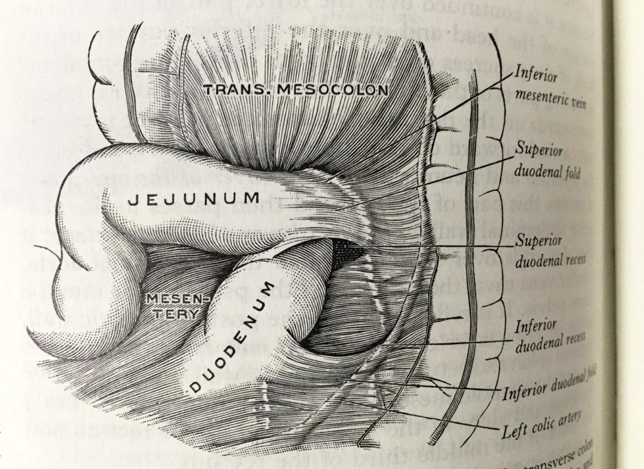
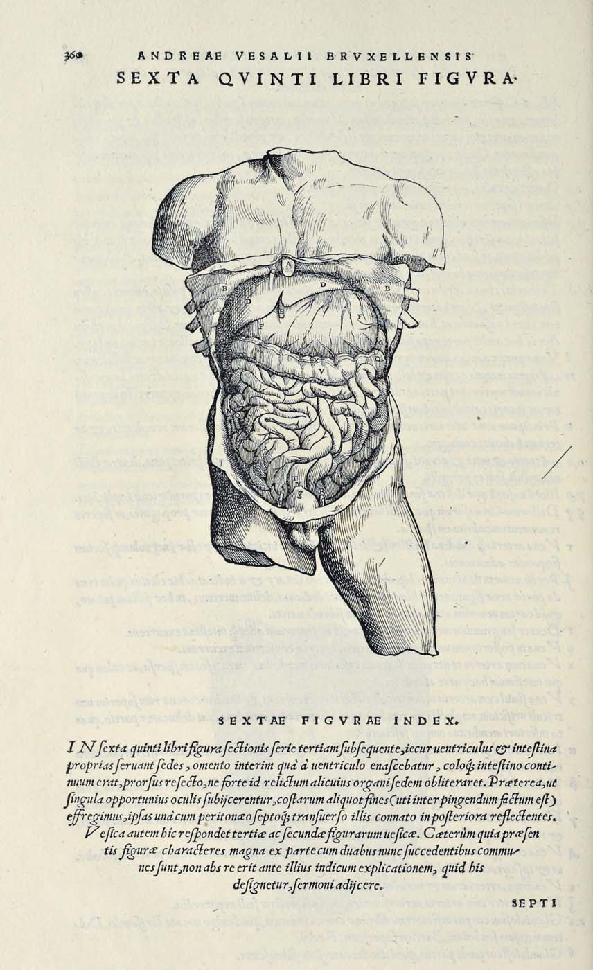
Henry Gray (anatomist) and Henry Vandyke Carter (artist), Fig. 1005, Superior and inferior duodenal fossæ.
The Buddha’s Guts
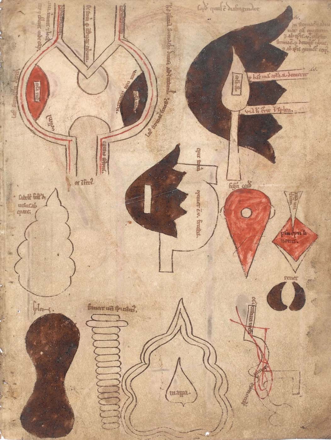
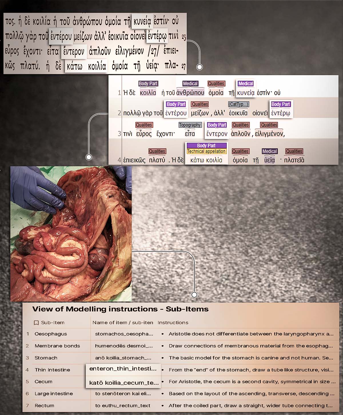
From Text to Model: The Guts according to Aristotle
Depiction of an eagle devouring a human heart at the site of Tula
Knitted artwork of guts based on Tibetan medical painting at Atsagat Monastery, Republic of Buryatia, Russia.
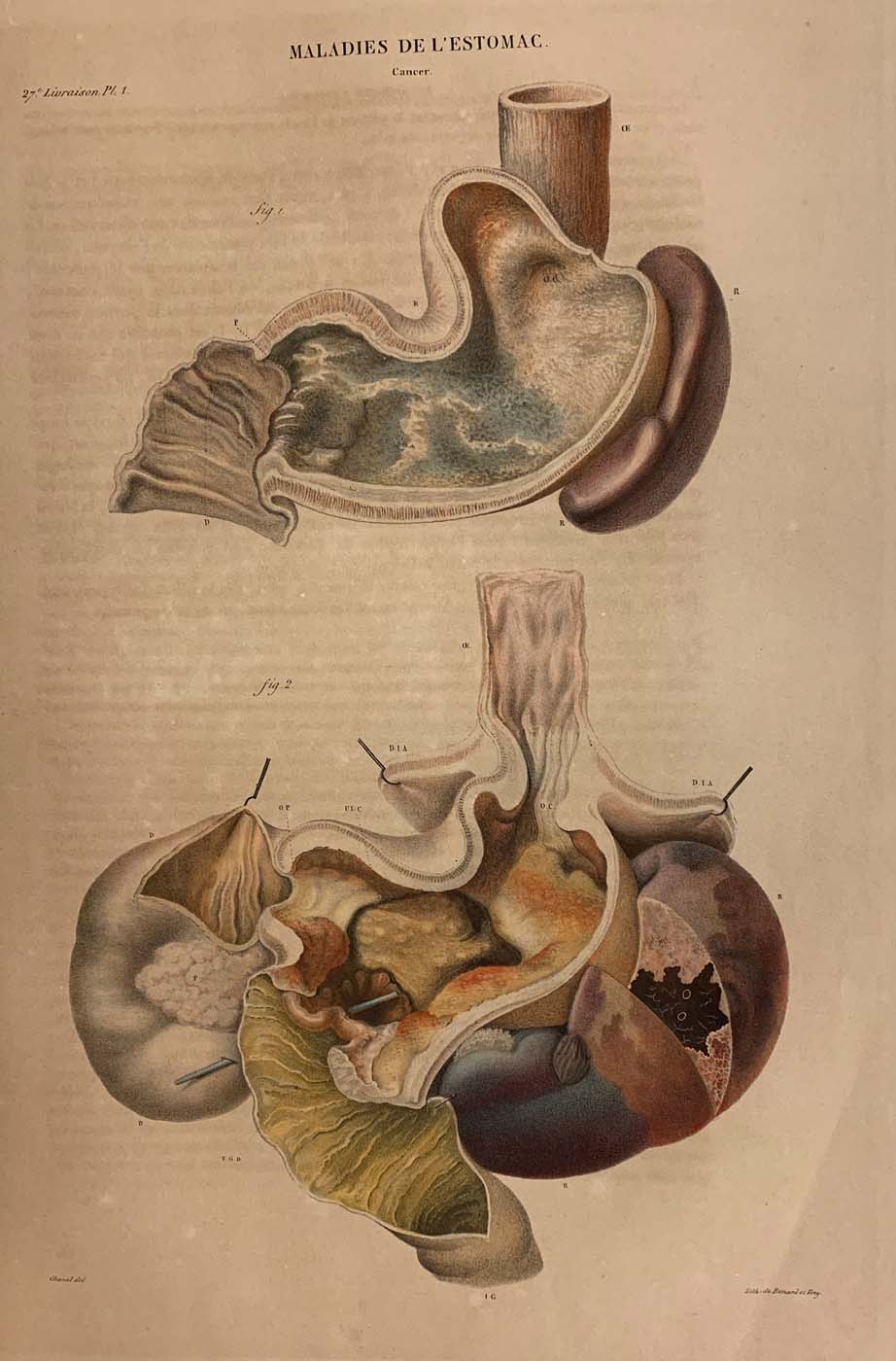
Diseases of the Stomach (cancer)
Major connecting blood vasculature of the body including gut with related vulnerable points
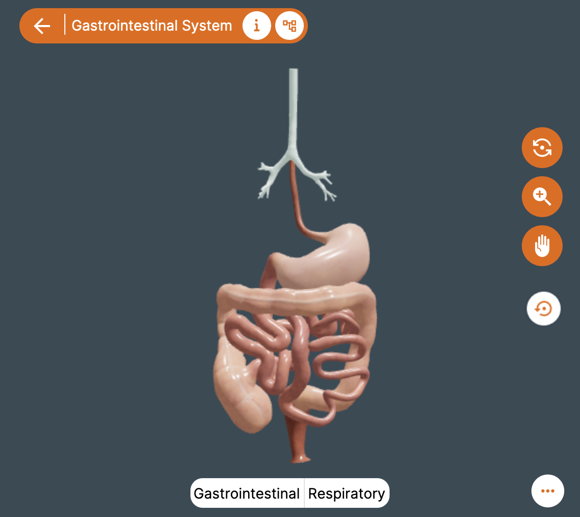
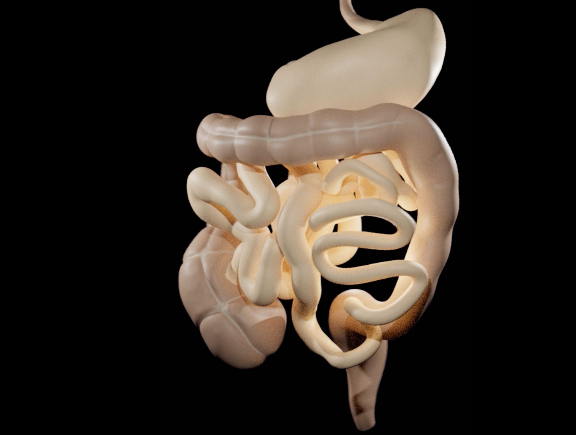
The 3D Model of the Digestive System according to Aristotle
Diagrams of the internal organs
Abdominal Dissection
Published in 1597 reprint of a 1565 Yixue gangmu, preserved in Ms sin. 11, part 1, “Enlightened-Hall Diagram of the Viscera”
A human anatomical figure
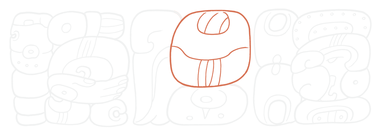
A segment of a Mayan hieroglyphic text, highlighting the stylised glyph for ‘heart’
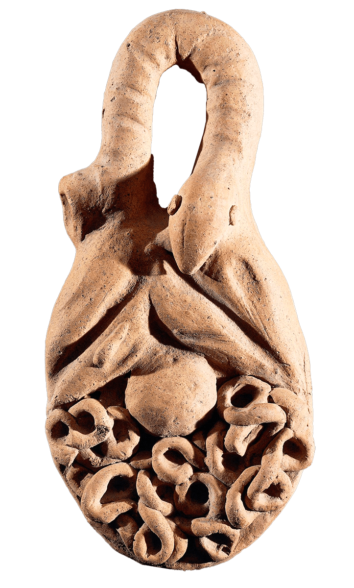
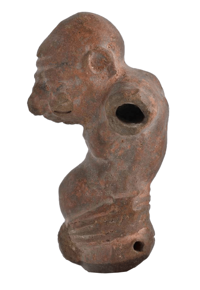
Grotesque figurine
Brain Frog
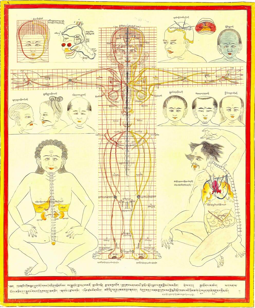
Maps of the inner sceneries • ventral and dorsal view
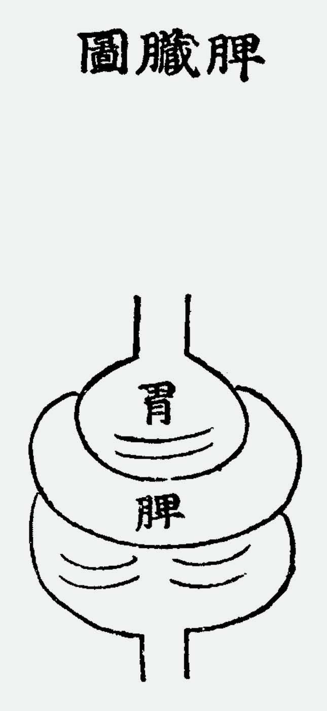
The Spleen and Stomach
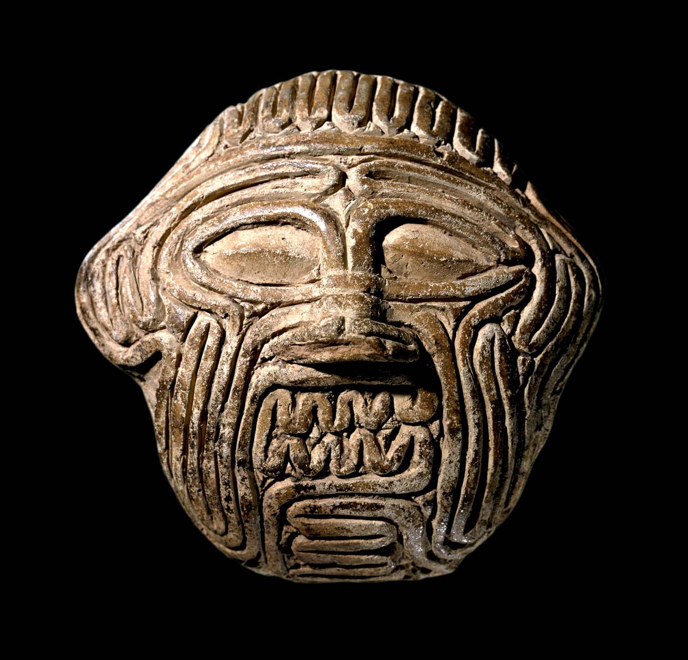
Mask of Huwawa
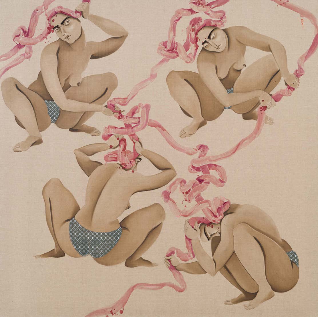
Side entanglements on Flax
Torshi and eyes
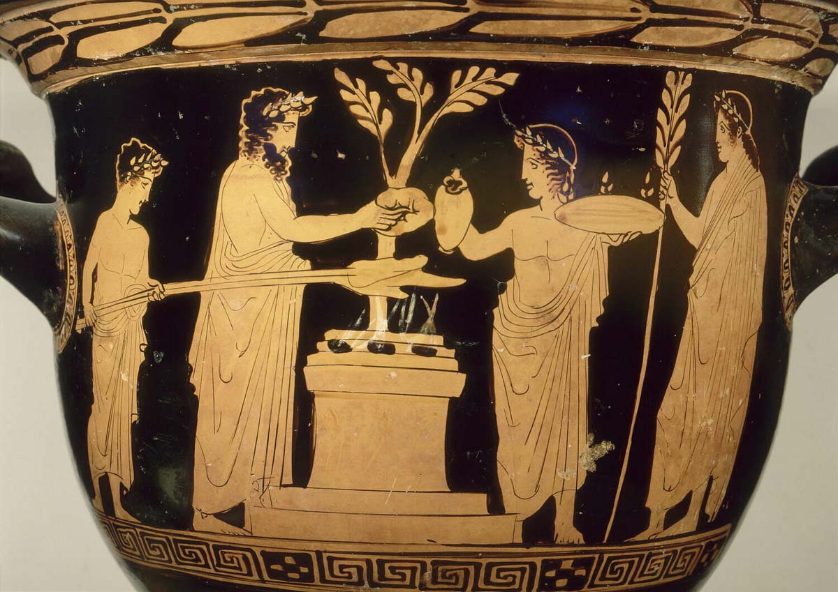
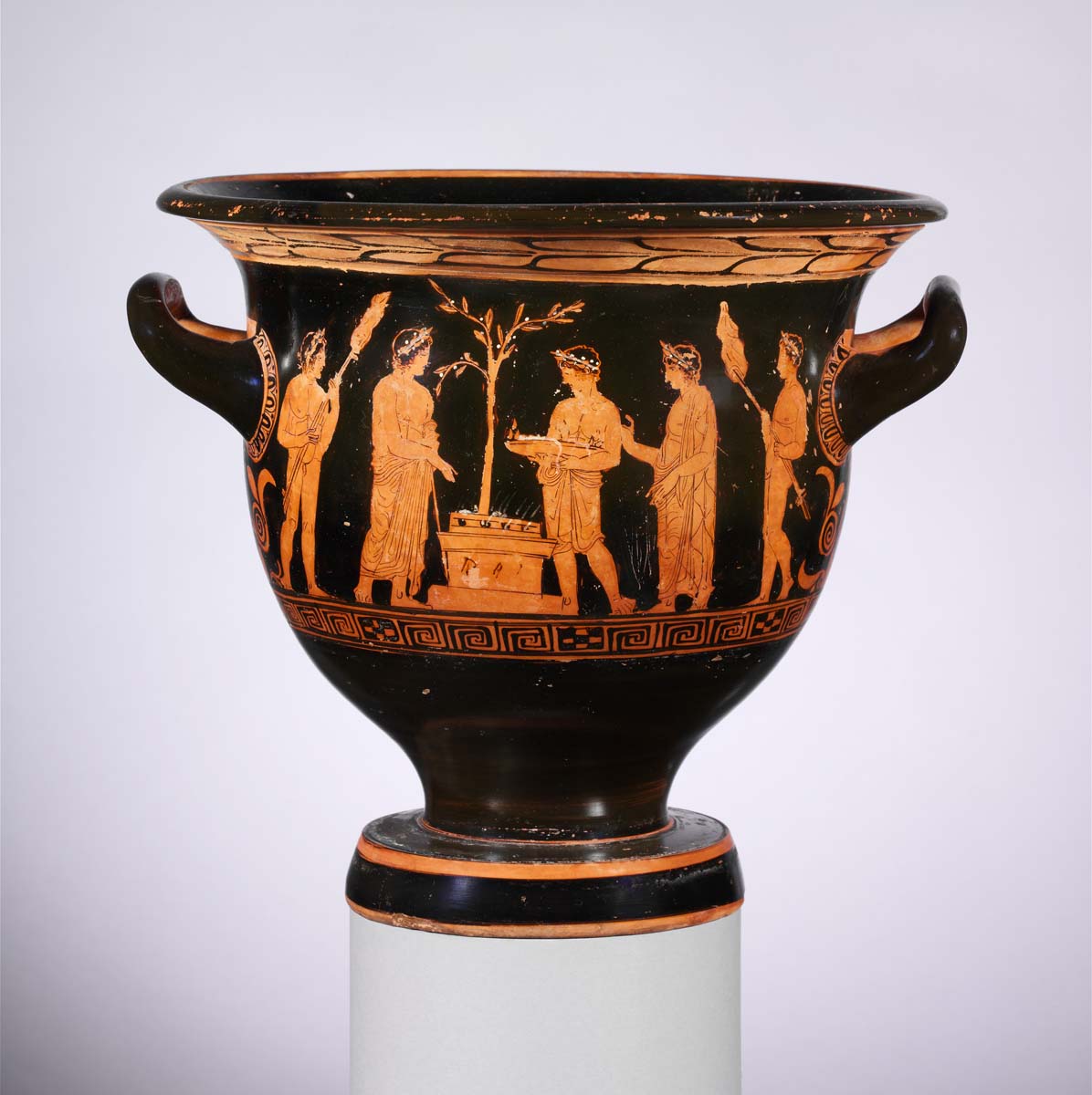
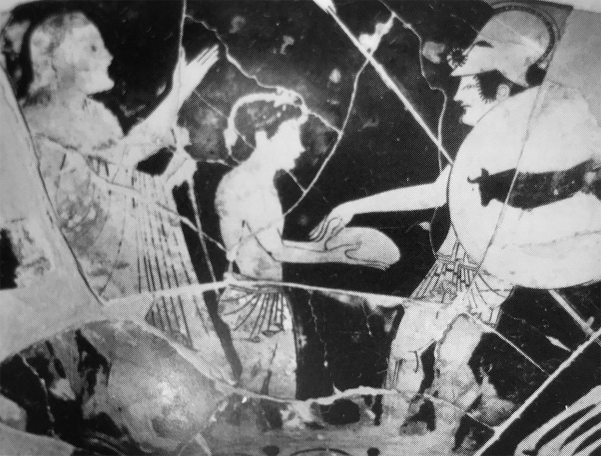
Athenian red-figure cup attributed to Oltos, examination of the liver of a sacrificial victim
The Gut / Der Darm
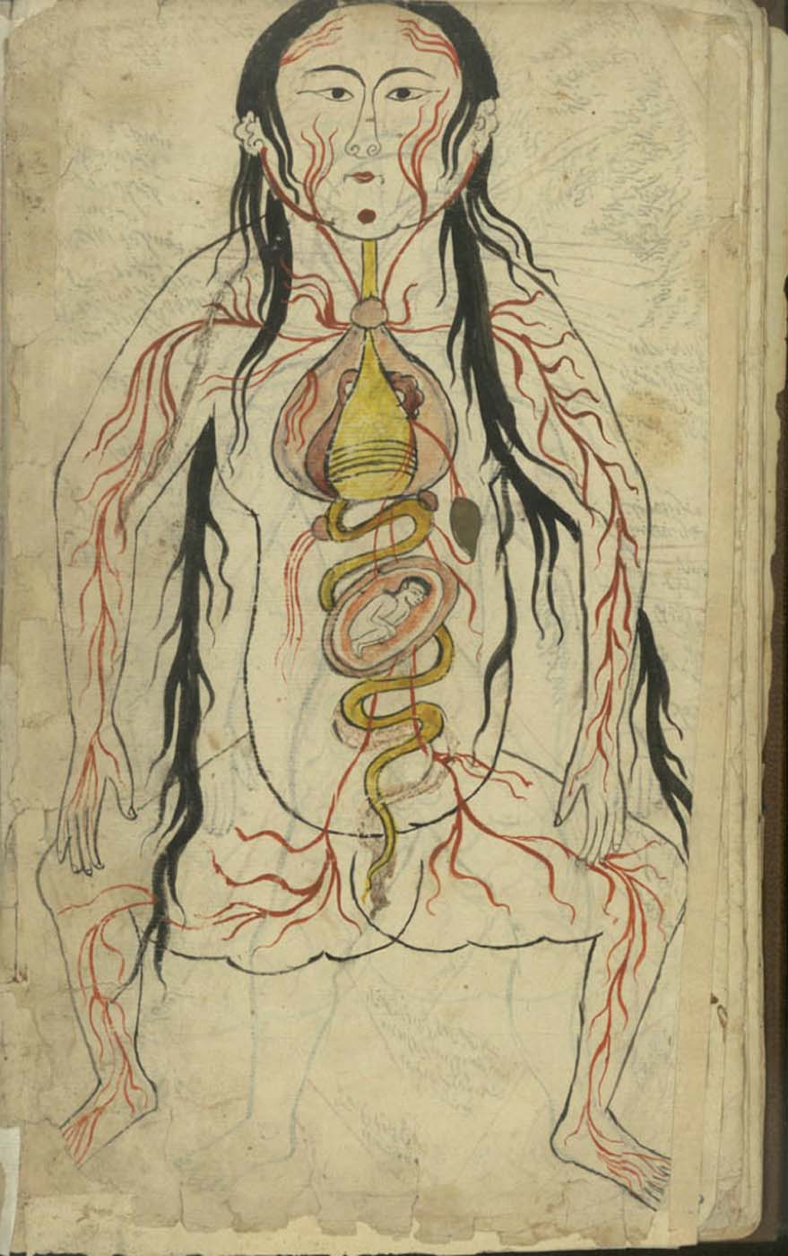
Representation of a woman with the distribution of the blood vessels and the internal organs (f. 15v)
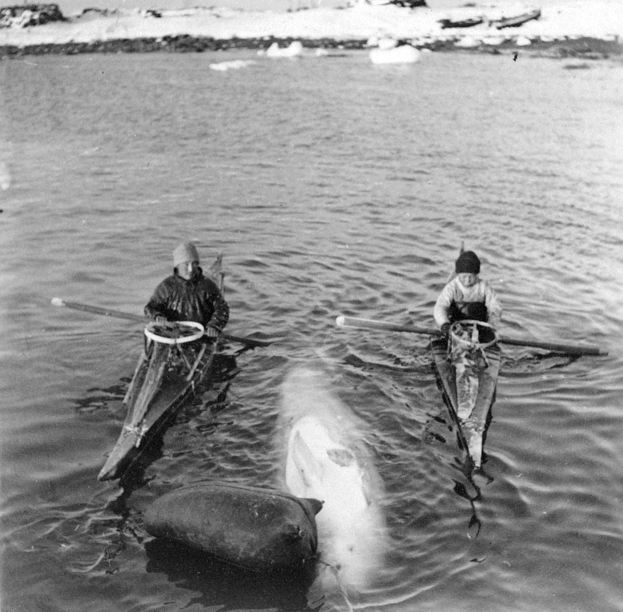
Towing bladder/hunting float.
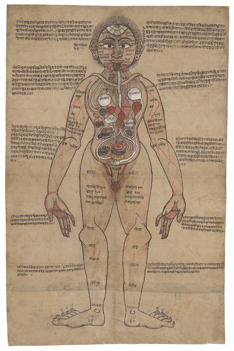
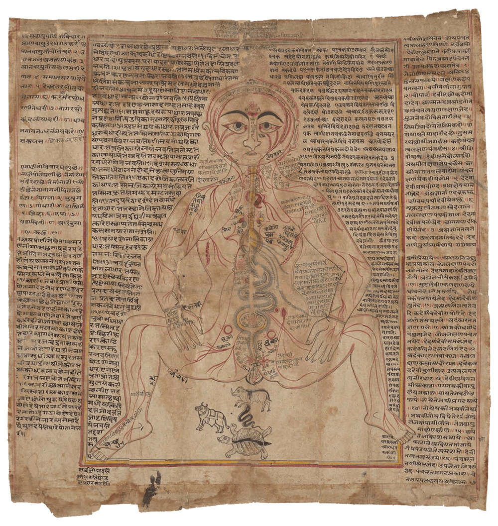
Indian anatomical painting
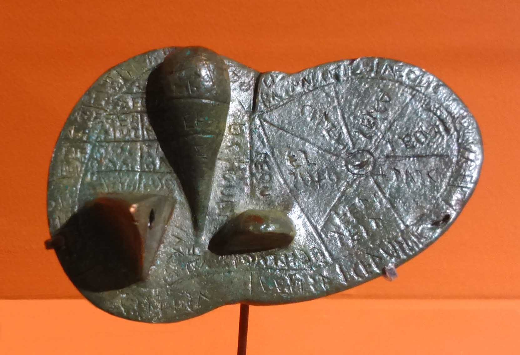
The ‘Piacenza Liver’
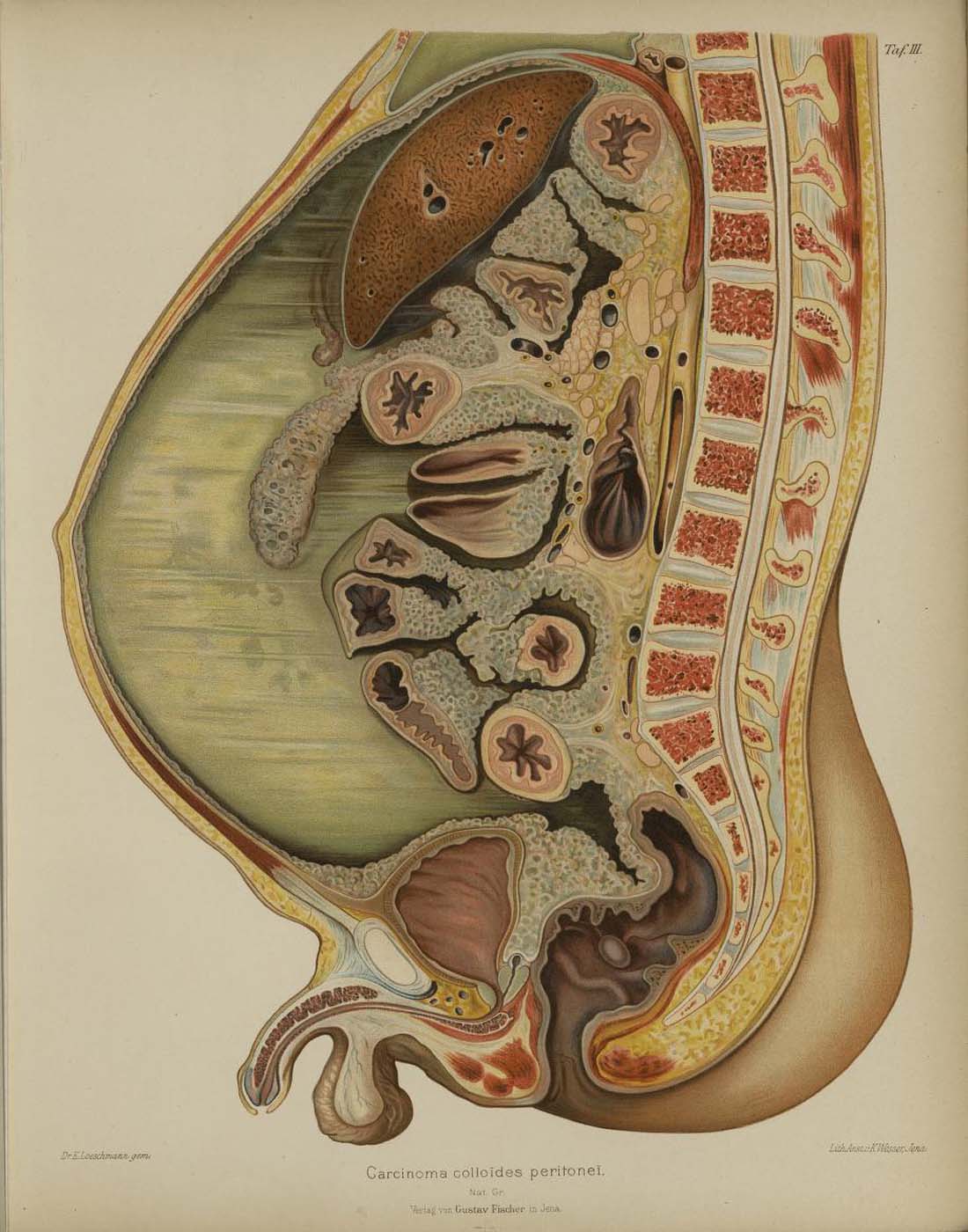
Carcinoma colloides peritonei
Load More
Viscera
Area
Histories
Image Descriptions
About